 Movie
Movie Controller


+ Open data

- Basic information
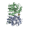
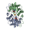
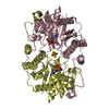
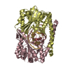
| Entry | Database: PDB / ID: 1g5p | ||||||
|---|---|---|---|---|---|---|---|
| Title | NITROGENASE IRON PROTEIN FROM AZOTOBACTER VINELANDII | ||||||
 Components Components | NITROGENASE IRON PROTEIN | ||||||
 Keywords Keywords | OXIDOREDUCTASE / IRON PROTEIN | ||||||
| Function / homology |  Function and homology information Function and homology informationnitrogenase / nitrogenase activity / 4 iron, 4 sulfur cluster binding / ATP binding / metal ion binding Similarity search - Function | ||||||
| Biological species |  Azotobacter vinelandii (bacteria) Azotobacter vinelandii (bacteria) | ||||||
| Method |  X-RAY DIFFRACTION / SYNCHROTRON / MOLECULAR REPLACEMENT / Resolution: 2.2 Å X-RAY DIFFRACTION / SYNCHROTRON / MOLECULAR REPLACEMENT / Resolution: 2.2 Å | ||||||
 Authors Authors | Strop, P. / Takahara, P.M. / Chiu, H.J. / Angove, H.C. / Burgess, B.K. / Rees, D.C. | ||||||
 Citation Citation | Journal: Biochemistry / Year: 2001 Title: Crystal structure of the all-ferrous [4Fe-4S]0 form of the nitrogenase iron protein from Azotobacter vinelandii. Authors: Strop, P. / Takahara, P.M. / Chiu, H. / Angove, H.C. / Burgess, B.K. / Rees, D.C. #1: Journal: Science / Year: 1992Title: Crystallographic Structure of the Nitrogenase Iron Protein from Azotobacter vinelandii Authors: KOMIYA, H. / GEORGIADIS, M.M. / CHAKRABARTI, P.P. / WOO, D. / KORNUC, J.J. / REES, D.C. #2: Journal: J.Mol.Biol. / Year: 1998Title: CONFORMATIONAL VARIABILITY IN STRUCTURES OF THE NITROGENASE IRON PROTEINS FROM AZOTOBACTER VINELANDII AND CLOSTRIDIUM PASTEURIANUM Authors: SCHLESSMAN, J.L. / WOO, D. / JOSHUA-TOR, L. / HOWARD, J.B. / REES, D.C. | ||||||
| History |
|
- Structure visualization
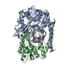
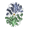
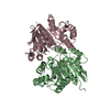
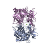
| Structure viewer | Molecule: MolmilJmol/JSmol |
|---|
- Downloads & links
-Download
| PDBx/mmCIF format | 1g5p.cif.gz | 127.4 KB | Display | PDBx/mmCIF format |
|---|---|---|---|---|
| PDB format | pdb1g5p.ent.gz | 98.3 KB | Display | PDB format |
| PDBx/mmJSON format | 1g5p.json.gz | Tree view | PDBx/mmJSON format | |
| Others |  Other downloads Other downloads |
-Validation report
| Arichive directory | https://data.pdbj.org/pub/pdb/validation_reports/g5/1g5pftp://data.pdbj.org/pub/pdb/validation_reports/g5/1g5p | HTTPS FTP |
|---|
-Related structure data
| Related structure data |  1g1mC  1nipS C: citing same article ( S: Starting model for refinement |
|---|---|
| Similar structure data |


-Links
 PDBj
PDBj
- Assembly
| Deposited unit | 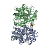
| ||||||||
|---|---|---|---|---|---|---|---|---|---|
| 1 |
| ||||||||
| Unit cell |
|
-Components
| #1: Protein | Mass: 31417.045 Da / Num. of mol.: 2 / Source method: isolated from a natural source / Details: GENE: NIFH / Source: (natural) Azotobacter vinelandii (bacteria) / Strain: OP / References: UniProt: P00459, nitrogenase#2: Chemical | ChemComp-SF4 / |   Mass: 351.640 Da / Num. of mol.: 1 / Source method: obtained synthetically / Formula: Fe4S4 Mass: 351.640 Da / Num. of mol.: 1 / Source method: obtained synthetically / Formula: Fe4S4#3: Water | ChemComp-HOH / |  Mass: 18.015 Da / Num. of mol.: 351 / Source method: isolated from a natural source / Formula: H2O Mass: 18.015 Da / Num. of mol.: 351 / Source method: isolated from a natural source / Formula: H2O |
|---|
-Experimental details
-Experiment
| Experiment | Method: X-RAY DIFFRACTION / Number of used crystals: 1 |
|---|
- Sample preparation
| Crystal | Density Matthews: 2.4 Å3/Da / Density % sol: 48.73 % | ||||||||||||||||||||||||||||||||||||||||||||||||
|---|---|---|---|---|---|---|---|---|---|---|---|---|---|---|---|---|---|---|---|---|---|---|---|---|---|---|---|---|---|---|---|---|---|---|---|---|---|---|---|---|---|---|---|---|---|---|---|---|---|
| Crystal grow | Temperature: 298 K / Method: liquid diffusion / pH: 8 Details: 25-30% PEG 4000, 140-200 mM NA2MOO4, 100 mM TRIS-CL, PH 8.0; PROTEIN WAS IN 20% GLYCEROL, 50 mM TRIS-CL, PH 8.0, 450 mM NACL, AND 2 mM NA2S2O4 , LIQUID DIFFUSION, temperature 298K | ||||||||||||||||||||||||||||||||||||||||||||||||
| Crystal grow | *PLUS Method: batch method | ||||||||||||||||||||||||||||||||||||||||||||||||
| Components of the solutions | *PLUS
|
-Data collection
| Diffraction | Mean temperature: 113 K |
|---|---|
| Diffraction source | Source: SYNCHROTRON / Site: SSRL  / Beamline: BL7-1 / Wavelength: 1.08 Å / Beamline: BL7-1 / Wavelength: 1.08 Å |
| Detector | Type: MARRESEARCH / Detector: IMAGE PLATE / Date: Mar 1, 1995 |
| Radiation | Monochromator: SI(111) / Protocol: SINGLE WAVELENGTH / Monochromatic (M) / Laue (L): M / Scattering type: x-ray |
| Radiation wavelength | Wavelength: 1.08 Å / Relative weight: 1 |
| Reflection | Resolution: 2.13→50 Å / Num. obs: 29060 / % possible obs: 92.9 % / Redundancy: 4 % / Rmerge(I) obs: 0.085 / Rsym value: 0.085 / Net I/σ(I): 6.9 |
| Reflection shell | Resolution: 2.2→2.28 Å / Redundancy: 3.8 % / Rmerge(I) obs: 0.309 / Mean I/σ(I) obs: 2.4 / Rsym value: 0.309 / % possible all: 94.9 |
- Processing
| Software |
| ||||||||||||||||
|---|---|---|---|---|---|---|---|---|---|---|---|---|---|---|---|---|---|
| Refinement | Method to determine structure: MOLECULAR REPLACEMENT Starting model: 1NIP Resolution: 2.2→50 Å / Cross valid method: THROUGHOUT / Stereochemistry target values: Engh & Huber
| ||||||||||||||||
| Refinement step | Cycle: LAST / Resolution: 2.2→50 Å
| ||||||||||||||||
| Refine LS restraints |
| ||||||||||||||||
| LS refinement shell | Resolution: 2.2→2.28 Å / Rfactor Rfree error: 0.025
| ||||||||||||||||
| Software | *PLUS Name: X-PLOR / Version: 3.851 / Classification: refinement | ||||||||||||||||
| Refinement | *PLUS Highest resolution: 2.2 Å / Lowest resolution: 50 Å / Rfactor Rfree: 0.29 | ||||||||||||||||
| Solvent computation | *PLUS | ||||||||||||||||
| Displacement parameters | *PLUS | ||||||||||||||||
| Refine LS restraints | *PLUS
| ||||||||||||||||
| LS refinement shell | *PLUS Highest resolution: 2.2 Å / Rfactor Rfree: 0.336 / Rfactor Rwork: 0.295 |
