 Movie
Movie Controller


+ Open data

- Basic information
| Entry | Database: PDB / ID: 1tw8 | ||||||
|---|---|---|---|---|---|---|---|
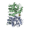
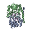
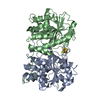
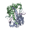
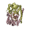
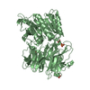
| Title | HincII bound to Ca2+ and cognate DNA GTCGAC | ||||||
 Components Components |
| ||||||
 Keywords Keywords | hydrolase/DNA / restriction endonuclease / hydrolase-DNA COMPLEX | ||||||
| Function / homology |  Function and homology information Function and homology informationtype II site-specific deoxyribonuclease / type II site-specific deoxyribonuclease activity / DNA restriction-modification system / DNA binding Similarity search - Function | ||||||
| Biological species |  Haemophilus influenzae (bacteria) Haemophilus influenzae (bacteria) | ||||||
| Method |  X-RAY DIFFRACTION / SYNCHROTRON / MOLECULAR REPLACEMENT / Resolution: 2.8 Å X-RAY DIFFRACTION / SYNCHROTRON / MOLECULAR REPLACEMENT / Resolution: 2.8 Å | ||||||
 Authors Authors | Etzkorn, C. / Horton, N.C. | ||||||
 Citation Citation | Journal: Biochemistry / Year: 2004 Title: Ca2+ binding in the active site of HincII: implications for the catalytic mechanism Authors: Etzkorn, C. / Horton, N.C. | ||||||
| History |
|
- Structure visualization
| Structure viewer | Molecule: MolmilJmol/JSmol |
|---|
- Downloads & links
-Download
| PDBx/mmCIF format | 1tw8.cif.gz | 268.7 KB | Display | PDBx/mmCIF format |
|---|---|---|---|---|
| PDB format | pdb1tw8.ent.gz | 210.5 KB | Display | PDB format |
| PDBx/mmJSON format | 1tw8.json.gz | Tree view | PDBx/mmJSON format | |
| Others |  Other downloads Other downloads |
-Validation report
| Arichive directory | https://data.pdbj.org/pub/pdb/validation_reports/tw/1tw8ftp://data.pdbj.org/pub/pdb/validation_reports/tw/1tw8 | HTTPS FTP |
|---|
-Related structure data
| Related structure data | 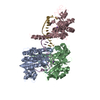 1tx3C  1kc6S S: Starting model for refinement C: citing same article ( |
|---|---|
| Similar structure data |


-Links
 PDBj
PDBj


- Assembly
| Deposited unit | 
| ||||||||
|---|---|---|---|---|---|---|---|---|---|
| 1 | 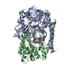
| ||||||||
| 2 | 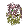
| ||||||||
| Unit cell |
| ||||||||
| Details | The biological assembly is one half of the asymmetric unit including two protein subunits and two strands of DNA (ie. chains A-B, E-F) |
-Components
| #1: DNA chain | Mass: 3993.586 Da / Num. of mol.: 4 / Source method: obtained synthetically / Details: commercial phosphoramidite chemistry #2: Protein | Mass: 29820.066 Da / Num. of mol.: 4 Source method: isolated from a genetically manipulated source Source: (gene. exp.) Haemophilus influenzae (bacteria) / Gene: Hinc II / Production host: References: UniProt: E1B6R0, UniProt: P17743*PLUS, type II site-specific deoxyribonuclease #3: Chemical | ChemComp-CA /   Mass: 40.078 Da / Num. of mol.: 6 / Source method: obtained synthetically / Formula: Ca Mass: 40.078 Da / Num. of mol.: 6 / Source method: obtained synthetically / Formula: Ca#4: Chemical | ChemComp-NA /   Mass: 22.990 Da / Num. of mol.: 4 / Source method: obtained synthetically / Formula: Na Mass: 22.990 Da / Num. of mol.: 4 / Source method: obtained synthetically / Formula: Na#5: Water | ChemComp-HOH / |  Mass: 18.015 Da / Num. of mol.: 765 / Source method: isolated from a natural source / Formula: H2O Mass: 18.015 Da / Num. of mol.: 765 / Source method: isolated from a natural source / Formula: H2O |
|---|
-Experimental details
-Experiment
| Experiment | Method: X-RAY DIFFRACTION / Number of used crystals: 1 |
|---|
- Sample preparation
| Crystal | Density Matthews: 2.79 Å3/Da / Density % sol: 55.98 % |
|---|---|
| Crystal grow | Temperature: 290 K / Method: vapor diffusion, hanging drop / pH: 5.5 Details: PEG 4000, 0.1M citrate, 0.15M NaCl, pH 5.5, VAPOR DIFFUSION, HANGING DROP, temperature 290K |
-Data collection
| Diffraction | Mean temperature: 100 K |
|---|---|
| Diffraction source | Source: SYNCHROTRON / Site: SSRL  / Beamline: BL9-1 / Wavelength: 0.9 Å / Beamline: BL9-1 / Wavelength: 0.9 Å |
| Detector | Type: ADSC QUANTUM 4 / Detector: CCD |
| Radiation | Protocol: SINGLE WAVELENGTH / Monochromatic (M) / Laue (L): M / Scattering type: x-ray |
| Radiation wavelength | Wavelength: 0.9 Å / Relative weight: 1 |
| Reflection | Resolution: 2.8→20 Å / Num. obs: 36795 / % possible obs: 96.9 % / Observed criterion σ(F): 2 / Observed criterion σ(I): 2 / Redundancy: 2.7 % / Biso Wilson estimate: 55.1 Å2 / Rmerge(I) obs: 0.066 / Rsym value: 0.066 / Net I/σ(I): 5.3 |
- Processing
| Software |
| |||||||||||||||||||||||||
|---|---|---|---|---|---|---|---|---|---|---|---|---|---|---|---|---|---|---|---|---|---|---|---|---|---|---|
| Refinement | Method to determine structure: MOLECULAR REPLACEMENT Starting model: PDB ENTRY 1KC6 Resolution: 2.8→20 Å / Cross valid method: Rfree / σ(F): 2 / σ(I): 2 / Stereochemistry target values: CNS
| |||||||||||||||||||||||||
| Refinement step | Cycle: LAST / Resolution: 2.8→20 Å
| |||||||||||||||||||||||||
| Refine LS restraints |
|
