 Movie
Movie Controller

[English] 日本語
 Yorodumi
- PDB-2gig: Alteration of sequence specificity of the type II restriction end... -
+ Open data

- Basic information
| Entry | Database: PDB / ID: 2gig | ||||||
|---|---|---|---|---|---|---|---|
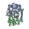
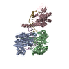
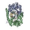
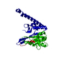
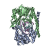
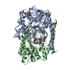
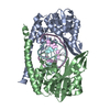
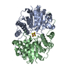
| Title | Alteration of sequence specificity of the type II restriction endonuclease HINCII through an indirect readout mechanism | ||||||
 Components Components |
| ||||||
 Keywords Keywords | HYDROLASE/DNA / Protein DNA complex / indirect readout / DNA intercalation / endonuclease / HYDROLASE-DNA COMPLEX | ||||||
| Function / homology |  Function and homology information Function and homology informationtype II site-specific deoxyribonuclease / type II site-specific deoxyribonuclease activity / DNA restriction-modification system / DNA binding Similarity search - Function | ||||||
| Biological species |  Haemophilus influenzae (bacteria) Haemophilus influenzae (bacteria) | ||||||
| Method |  X-RAY DIFFRACTION / SYNCHROTRON / MOLECULAR REPLACEMENT / Resolution: 1.83 Å X-RAY DIFFRACTION / SYNCHROTRON / MOLECULAR REPLACEMENT / Resolution: 1.83 Å | ||||||
 Authors Authors | Horton, N.C. / Joshi, H.K. / Etzkorn, C. / Chatwell, L. / Bitinaite, J. | ||||||
 Citation Citation | Journal: J.Biol.Chem. / Year: 2006 Title: Alteration of Sequence Specificity of the Type II Restriction Endonuclease HincII through an Indirect Readout Mechanism. Authors: Joshi, H.K. / Etzkorn, C. / Chatwell, L. / Bitinaite, J. / Horton, N.C. | ||||||
| History |
|
- Structure visualization
| Structure viewer | Molecule: MolmilJmol/JSmol |
|---|
- Downloads & links
-Download
| PDBx/mmCIF format | 2gig.cif.gz | 140.3 KB | Display | PDBx/mmCIF format |
|---|---|---|---|---|
| PDB format | pdb2gig.ent.gz | 104.8 KB | Display | PDB format |
| PDBx/mmJSON format | 2gig.json.gz | Tree view | PDBx/mmJSON format | |
| Others |  Other downloads Other downloads |
-Validation report
| Arichive directory | https://data.pdbj.org/pub/pdb/validation_reports/gi/2gigftp://data.pdbj.org/pub/pdb/validation_reports/gi/2gig | HTTPS FTP |
|---|
-Related structure data
| Related structure data | 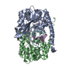 2gieC 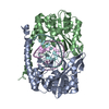 2gihC 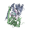 2giiC 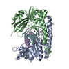 2gijC 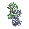 1tw3S C: citing same article ( S: Starting model for refinement |
|---|---|
| Similar structure data |


-Links
 PDBj
PDBj


- Assembly
| Deposited unit | 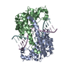
| ||||||||
|---|---|---|---|---|---|---|---|---|---|
| 1 |
| ||||||||
| Unit cell |
|
-Components
| #1: DNA chain | Mass: 4282.767 Da / Num. of mol.: 2 / Source method: obtained synthetically #2: Protein | Mass: 29839.109 Da / Num. of mol.: 2 / Mutation: Q138F Source method: isolated from a genetically manipulated source Source: (gene. exp.) Haemophilus influenzae (bacteria) / Gene: hincIIR / Plasmid: pCCR101HincII.R / Production host: References: UniProt: P44413, UniProt: P17743*PLUS, type II site-specific deoxyribonuclease #3: Chemical |   Mass: 22.990 Da / Num. of mol.: 2 / Source method: obtained synthetically / Formula: Na Mass: 22.990 Da / Num. of mol.: 2 / Source method: obtained synthetically / Formula: Na#4: Water | ChemComp-HOH / |  Mass: 18.015 Da / Num. of mol.: 463 / Source method: isolated from a natural source / Formula: H2O Mass: 18.015 Da / Num. of mol.: 463 / Source method: isolated from a natural source / Formula: H2O |
|---|
-Experimental details
-Experiment
| Experiment | Method: X-RAY DIFFRACTION / Number of used crystals: 1 |
|---|
- Sample preparation
| Crystal | Density Matthews: 2.18 Å3/Da / Density % sol: 43.61 % | ||||||||||||||||||||||||||||||||||||
|---|---|---|---|---|---|---|---|---|---|---|---|---|---|---|---|---|---|---|---|---|---|---|---|---|---|---|---|---|---|---|---|---|---|---|---|---|---|
| Crystal grow | Temperature: 290 K / Method: vapor diffusion, hanging drop / pH: 7.5 Details: 25% PEG 4K, 0.15 M NaCl, 0.1 M HEPES, pH 7.5, VAPOR DIFFUSION, HANGING DROP, temperature 290K | ||||||||||||||||||||||||||||||||||||
| Components of the solutions |
|
-Data collection
| Diffraction | Mean temperature: 100 K |
|---|---|
| Diffraction source | Source: SYNCHROTRON / Site: SSRL  / Beamline: BL9-2 / Wavelength: 0.9 Å / Beamline: BL9-2 / Wavelength: 0.9 Å |
| Detector | Type: ADSC QUANTUM 4 / Detector: CCD / Date: Jul 30, 2004 |
| Radiation | Monochromator: 180 frames, 1 degree each, 30 sec per frame / Protocol: SINGLE WAVELENGTH / Monochromatic (M) / Laue (L): M / Scattering type: x-ray |
| Radiation wavelength | Wavelength: 0.9 Å / Relative weight: 1 |
| Reflection | Resolution: 1.83→50 Å / Num. obs: 47047 / % possible obs: 90.7 % / Observed criterion σ(F): -3 / Observed criterion σ(I): -3 / Redundancy: 3.6 % / Biso Wilson estimate: 26.7 Å2 / Rmerge(I) obs: 0.046 / Net I/σ(I): 9.1 |
| Reflection shell | Resolution: 1.83→1.88 Å / Redundancy: 3.3 % / Rmerge(I) obs: 0.318 / Mean I/σ(I) obs: 2.1 / % possible all: 90.7 |
- Processing
| Software |
| |||||||||||||||||||||||||
|---|---|---|---|---|---|---|---|---|---|---|---|---|---|---|---|---|---|---|---|---|---|---|---|---|---|---|
| Refinement | Method to determine structure: MOLECULAR REPLACEMENT Starting model: pdb entry 1TW3 Resolution: 1.83→50 Å / σ(F): 2 / Stereochemistry target values: Engh & Huber
| |||||||||||||||||||||||||
| Refinement step | Cycle: LAST / Resolution: 1.83→50 Å
| |||||||||||||||||||||||||
| Refine LS restraints |
|
