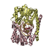
 Movie
Movie Controller


+ Open data

- Basic information
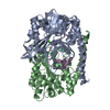
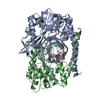
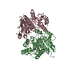
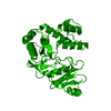
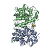
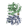
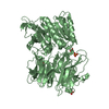
| Entry | Database: PDB / ID: 1xhv | ||||||
|---|---|---|---|---|---|---|---|
| Title | HincII bound to cleaved cognate DNA GTCGAC and Mn2+ | ||||||
 Components Components |
| ||||||
 Keywords Keywords | hydrolase/DNA / restriction endonuclease / phosphoryl transfer / hydrolase-DNA COMPLEX | ||||||
| Function / homology |  Function and homology information Function and homology informationtype II site-specific deoxyribonuclease / type II site-specific deoxyribonuclease activity / DNA restriction-modification system / DNA binding Similarity search - Function | ||||||
| Biological species |  Haemophilus influenzae (bacteria) Haemophilus influenzae (bacteria) | ||||||
| Method |  X-RAY DIFFRACTION / SYNCHROTRON / MOLECULAR REPLACEMENT / Resolution: 2.5 Å X-RAY DIFFRACTION / SYNCHROTRON / MOLECULAR REPLACEMENT / Resolution: 2.5 Å | ||||||
 Authors Authors | Etzkorn, C. / Horton, N.C. | ||||||
 Citation Citation | Journal: J.Mol.Biol. / Year: 2004 Title: Mechanistic Insights from the Structures of HincII Bound to Cognate DNA Cleaved from Addition of Mg(2+) and Mn(2+) Authors: Etzkorn, C. / Horton, N.C. | ||||||
| History |
|
- Structure visualization
| Structure viewer | Molecule: MolmilJmol/JSmol |
|---|
- Downloads & links
-Download
| PDBx/mmCIF format | 1xhv.cif.gz | 273.8 KB | Display | PDBx/mmCIF format |
|---|---|---|---|---|
| PDB format | pdb1xhv.ent.gz | 213.8 KB | Display | PDB format |
| PDBx/mmJSON format | 1xhv.json.gz | Tree view | PDBx/mmJSON format | |
| Others |  Other downloads Other downloads |
-Validation report
| Arichive directory | https://data.pdbj.org/pub/pdb/validation_reports/xh/1xhvftp://data.pdbj.org/pub/pdb/validation_reports/xh/1xhv | HTTPS FTP |
|---|
-Related structure data
| Related structure data |  1xhuC 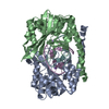 1kc6S S: Starting model for refinement C: citing same article ( |
|---|---|
| Similar structure data |


-Links
 PDBj
PDBj


- Assembly
| Deposited unit | 
| ||||||||
|---|---|---|---|---|---|---|---|---|---|
| 1 | 
| ||||||||
| 2 | 
| ||||||||
| Unit cell |
| ||||||||
| Components on special symmetry positions |
|
-Components
| #1: DNA chain | Mass: 2114.398 Da / Num. of mol.: 4 / Source method: obtained synthetically / Details: phosphoramidite synthetic chemistry #2: DNA chain | Mass: 1834.230 Da / Num. of mol.: 4 / Source method: obtained synthetically / Details: phosphoramidite synthetic chemistry #3: Protein | Mass: 29820.066 Da / Num. of mol.: 4 Source method: isolated from a genetically manipulated source Source: (gene. exp.) Haemophilus influenzae (bacteria) / Gene: hincIIR / Production host: References: UniProt: P17743, type II site-specific deoxyribonuclease #4: Chemical | ChemComp-MN /   Mass: 54.938 Da / Num. of mol.: 22 / Source method: obtained synthetically / Formula: Mn Mass: 54.938 Da / Num. of mol.: 22 / Source method: obtained synthetically / Formula: Mn#5: Water | ChemComp-HOH / |  Mass: 18.015 Da / Num. of mol.: 1027 / Source method: isolated from a natural source / Formula: H2O Mass: 18.015 Da / Num. of mol.: 1027 / Source method: isolated from a natural source / Formula: H2O |
|---|
-Experimental details
-Experiment
| Experiment | Method: X-RAY DIFFRACTION / Number of used crystals: 1 |
|---|
- Sample preparation
| Crystal | Density Matthews: 2.83 Å3/Da / Density % sol: 56.6 % | ||||||||||||||||
|---|---|---|---|---|---|---|---|---|---|---|---|---|---|---|---|---|---|
| Crystal grow | Temperature: 290 K / Method: vapor diffusion, hanging drop / pH: 5.5 Details: PEG 4000, NaCl, citrate, pH 5.5, VAPOR DIFFUSION, HANGING DROP, temperature 290K | ||||||||||||||||
| Components of the solutions |
|
-Data collection
| Diffraction | Mean temperature: 100 K |
|---|---|
| Diffraction source | Source: SYNCHROTRON / Site: APS  / Beamline: 17-BM / Wavelength: 0.9 Å / Beamline: 17-BM / Wavelength: 0.9 Å |
| Radiation | Protocol: SINGLE WAVELENGTH / Monochromatic (M) / Laue (L): M / Scattering type: x-ray |
| Radiation wavelength | Wavelength: 0.9 Å / Relative weight: 1 |
| Reflection | Resolution: 2.5→50 Å / Num. obs: 53281 / % possible obs: 89.3 % / Observed criterion σ(F): -3 / Observed criterion σ(I): -3 / Redundancy: 3.3 % / Biso Wilson estimate: 58.3 Å2 / Rmerge(I) obs: 0.064 / Rsym value: 0.064 / Net I/σ(I): 16.2 |
- Processing
| Software |
| ||||||||||||||||||||
|---|---|---|---|---|---|---|---|---|---|---|---|---|---|---|---|---|---|---|---|---|---|
| Refinement | Method to determine structure: MOLECULAR REPLACEMENT Starting model: 1KC6 without solvent Resolution: 2.5→50 Å / Cross valid method: R free / σ(F): 2 / Stereochemistry target values: as in CNS
| ||||||||||||||||||||
| Refinement step | Cycle: LAST / Resolution: 2.5→50 Å
| ||||||||||||||||||||
| Refine LS restraints |
|
