HETEROGEN According to authors, the ethylene glycol units are intended to model partially defined ...HETEROGEN According to authors, the ethylene glycol units are intended to model partially defined PEG molecules. None of them is an actual compound. The PEG used in the setup was PEG 10,000. The sodium atom 2492 is associated with water molecules 5025, 5026, 5027, 5028, the calcium atom 2490 with water molecules 5021, 5022 and calcium atom 2491 with water molecules 5023, 5024.
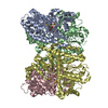
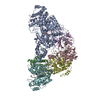
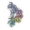
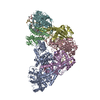
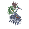
A: Nitrogenase molybdenum-iron protein B: Nitrogenase molybdenum-iron protein C: Nitrogenase molybdenum-iron protein D: Nitrogenase molybdenum-iron protein E: Nitrogenase iron protein 1 F: Nitrogenase iron protein 1 hetero molecules
In the structure databanks used in Yorodumi, some data are registered as the other names, "COVID-19 virus" and "2019-nCoV". Here are the details of the virus and the list of structure data.
Jan 31, 2019. EMDB accession codes are about to change! (news from PDBe EMDB page)
EMDB accession codes are about to change! (news from PDBe EMDB page)
The allocation of 4 digits for EMDB accession codes will soon come to an end. Whilst these codes will remain in use, new EMDB accession codes will include an additional digit and will expand incrementally as the available range of codes is exhausted. The current 4-digit format prefixed with “EMD-” (i.e. EMD-XXXX) will advance to a 5-digit format (i.e. EMD-XXXXX), and so on. It is currently estimated that the 4-digit codes will be depleted around Spring 2019, at which point the 5-digit format will come into force.
The EM Navigator/Yorodumi systems omit the EMD- prefix.
Related info.:Q: What is EMD? / ID/Accession-code notation in Yorodumi/EM Navigator
Yorodumi is a browser for structure data from EMDB, PDB, SASBDB, etc.
This page is also the successor to EM Navigator detail page, and also detail information page/front-end page for Omokage search.
The word "yorodu" (or yorozu) is an old Japanese word meaning "ten thousand". "mi" (miru) is to see.
Related info.:EMDB / PDB / SASBDB / Comparison of 3 databanks / Yorodumi Search / Aug 31, 2016. New EM Navigator & Yorodumi / Yorodumi Papers / Jmol/JSmol / Function and homology information / Changes in new EM Navigator and Yorodumi
 Movie
Movie Controller


 Open data
 Basic information
 Components
 Keywords
 Function and homology information
 Azotobacter vinelandii (bacteria)
 X-RAY DIFFRACTION /
  Authors
 Citation
 Structure visualization
 Downloads & links
 Other downloads


 PDBj
PDBj
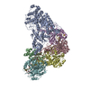
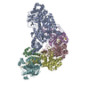
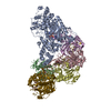
 Assembly
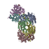
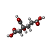


 Mass: 206.150 Da / Num. of mol.: 2 / Source method: obtained synthetically / Formula: C7H10O7
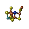
 Mass: 789.447 Da / Num. of mol.: 2 / Source method: obtained synthetically / Formula: Fe7MoNS9
 Mass: 150.173 Da / Num. of mol.: 8 / Source method: obtained synthetically / Formula: C6H14O4
 Mass: 40.078 Da / Num. of mol.: 2 / Source method: obtained synthetically / Formula: Ca
 Mass: 22.990 Da / Num. of mol.: 1 / Source method: obtained synthetically / Formula: Na
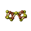
 Mass: 671.215 Da / Num. of mol.: 2 / Source method: obtained synthetically / Formula: Fe8S7
 Mass: 122.143 Da / Num. of mol.: 2 / Source method: obtained synthetically / Formula: C4H12NO3 / Comment: pH buffer*YM
 Mass: 194.226 Da / Num. of mol.: 2 / Source method: obtained synthetically / Formula: C8H18O5 / Comment: precipitant*YM
 Mass: 238.278 Da / Num. of mol.: 2 / Source method: obtained synthetically / Formula: C10H22O6 / Comment: precipitant*YM
 Mass: 106.120 Da / Num. of mol.: 3 / Source method: obtained synthetically / Formula: C4H10O3
 Mass: 282.331 Da / Num. of mol.: 1 / Source method: obtained synthetically / Formula: C12H26O7 / Comment: precipitant*YM
 Mass: 351.640 Da / Num. of mol.: 1 / Source method: obtained synthetically / Formula: Fe4S4
 Sample preparation
 / Beamline: BL9-1 / Wavelength: 0.9796 Å
 Processing