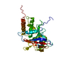
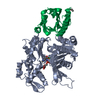
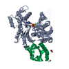
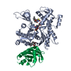
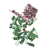
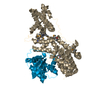
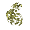
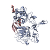
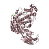
[English] 日本語
Yorodumi
- PDB-2b3o: Crystal structure of human tyrosine phosphatase SHP-1 -

+
Open data


ID or keywords:

Loading...

-
Basic information

Entry
Database: PDB / ID: 2b3o
TitleCrystal structure of human tyrosine phosphatase SHP-1
ComponentsTyrosine-protein phosphatase, non-receptor type 6
KeywordsHYDROLASE / Protein Tyrosine Phosphatase / SHP-1 / Signaling
Function / homology
Function and homology information


negative regulation of humoral immune response mediated by circulating immunoglobulin / negative regulation of mast cell activation involved in immune response / regulation of B cell differentiation / negative regulation of peptidyl-tyrosine phosphorylation / epididymis development / negative regulation of inflammatory response to wounding / phosphorylation-dependent protein binding / transmembrane receptor protein tyrosine phosphatase activity / natural killer cell mediated cytotoxicity / alpha-beta T cell receptor complex ...negative regulation of humoral immune response mediated by circulating immunoglobulin / negative regulation of mast cell activation involved in immune response / regulation of B cell differentiation / negative regulation of peptidyl-tyrosine phosphorylation / epididymis development / negative regulation of inflammatory response to wounding / phosphorylation-dependent protein binding / transmembrane receptor protein tyrosine phosphatase activity / natural killer cell mediated cytotoxicity / alpha-beta T cell receptor complex / regulation of release of sequestered calcium ion into cytosol / CD22 mediated BCR regulation / Interleukin-37 signaling / positive regulation of cell adhesion mediated by integrin / Costimulation by the CD28 family / Signal regulatory protein family interactions / platelet formation / negative regulation of T cell receptor signaling pathway / megakaryocyte development / negative regulation of MAPK cascade / Regulation of KIT signaling / Signaling by ALK / Platelet sensitization by LDL / regulation of G1/S transition of mitotic cell cycle / PECAM1 interactions / regulation of type I interferon-mediated signaling pathway / negative regulation of interleukin-6 production / negative regulation of tumor necrosis factor production / non-membrane spanning protein tyrosine phosphatase activity / peptidyl-tyrosine dephosphorylation / Interleukin-3, Interleukin-5 and GM-CSF signaling / Regulation of IFNA/IFNB signaling / PD-1 signaling / Interleukin receptor SHC signaling / hematopoietic progenitor cell differentiation / Nuclear events stimulated by ALK signaling in cancer / Regulation of IFNG signaling / T cell proliferation / negative regulation of T cell proliferation / Growth hormone receptor signaling / cell adhesion molecule binding / GPVI-mediated activation cascade / T cell costimulation / phosphotyrosine residue binding / SH2 domain binding / protein dephosphorylation / protein-tyrosine-phosphatase / regulation of ERK1 and ERK2 cascade / Antigen activates B Cell Receptor (BCR) leading to generation of second messengers / protein tyrosine phosphatase activity / B cell receptor signaling pathway / cytokine-mediated signaling pathway / platelet aggregation / SH3 domain binding / peptidyl-tyrosine phosphorylation / specific granule lumen / Interferon gamma signaling / MAPK cascade / Interferon alpha/beta signaling / cell-cell junction / tertiary granule lumen / mitotic cell cycle / T cell receptor signaling pathway / regulation of apoptotic process / positive regulation of phosphatidylinositol 3-kinase/protein kinase B signal transduction / cell differentiation / intracellular signal transduction / G protein-coupled receptor signaling pathway / negative regulation of cell population proliferation / positive regulation of cell population proliferation / Neutrophil degranulation / nucleolus / protein kinase binding / SARS-CoV-2 activates/modulates innate and adaptive immune responses / protein-containing complex / extracellular exosome / extracellular region / nucleoplasm / membrane / nucleus / cytoplasm / cytosol
Similarity search - Function
Protein-tyrosine phosphatase, non-receptor type-6, -11 / SH2 domain / SHC Adaptor Protein / Protein tyrosine phosphatase superfamily / Protein-Tyrosine Phosphatase; Chain A / Protein tyrosine phosphatase, catalytic domain / PTP type protein phosphatase domain profile. / Protein-tyrosine phosphatase / Tyrosine-specific protein phosphatase, PTPase domain / Protein-tyrosine phosphatase, catalytic ...Protein-tyrosine phosphatase, non-receptor type-6, -11 / SH2 domain / SHC Adaptor Protein / Protein tyrosine phosphatase superfamily / Protein-Tyrosine Phosphatase; Chain A / Protein tyrosine phosphatase, catalytic domain / PTP type protein phosphatase domain profile. / Protein-tyrosine phosphatase / Tyrosine-specific protein phosphatase, PTPase domain / Protein-tyrosine phosphatase, catalytic / Protein tyrosine phosphatase, catalytic domain motif / Tyrosine specific protein phosphatases active site. / Protein-tyrosine phosphatase, active site / Tyrosine-specific protein phosphatases domain / Tyrosine specific protein phosphatases domain profile. / Protein-tyrosine phosphatase-like / SH2 domain / Src homology 2 (SH2) domain profile. / Src homology 2 domains / SH2 domain / SH2 domain superfamily / Alpha-Beta Complex / 2-Layer Sandwich / Alpha Beta
Similarity search - Domain/homology
Tyrosine-protein phosphatase non-receptor type 6
Similarity search - Component
Biological speciesHomo sapiens (human)
MethodX-RAY DIFFRACTION / SYNCHROTRON / MOLECULAR REPLACEMENT / Resolution: 2.8 Å
AuthorsYang, J. / Liu, L. / He, D. / Song, X. / Liang, X. / Zhao, Z.J. / Zhou, G.W.
CitationJournal: J.Biol.Chem. / Year: 2003
Title: Crystal structure of human protein-tyrosine phosphatase SHP-1.
Authors: Yang, J. / Liu, L. / He, D. / Song, X. / Liang, X. / Zhao, Z.J. / Zhou, G.W.
History
DepositionSep 20, 2005Deposition site: RCSB / Processing site: RCSB
Revision 1.0Oct 25, 2005Provider: repository / Type: Initial release
Revision 1.1May 1, 2008Group: Version format compliance
Revision 1.2Jul 13, 2011Group: Version format compliance
Revision 1.3Feb 14, 2024Group: Data collection / Database references / Category: chem_comp_atom / chem_comp_bond / database_2
Item: _database_2.pdbx_DOI / _database_2.pdbx_database_accession

-
Structure visualization

Structure viewerMolecule:
MolmilJmol/JSmol

Downloads & links

-
Assembly

Deposited unit
A: Tyrosine-protein phosphatase, non-receptor type 6


Theoretical massNumber of molelcules
Total (without water)60,3511
Polymers60,3511
Non-polymers00
Water34219
1


  • Idetical with deposited unit
  • defined by author
TypeNameSymmetry operationNumber
identity operation1_555x,y,z1
Unit cell
Length a, b, c (Å)44.736, 100.404, 149.475
Angle α, β, γ (deg.)90.00, 90.00, 90.00
Int Tables number19
Space group name H-MP212121

-
Components

#1: Protein Tyrosine-protein phosphatase, non-receptor type 6 / Protein-tyrosine phosphatase 1C / PTP-1C / Hematopoietic cell protein-tyrosine phosphatase / SH- ...Protein-tyrosine phosphatase 1C / PTP-1C / Hematopoietic cell protein-tyrosine phosphatase / SH-PTP1 / Protein-tyrosine phosphatase SHP-1


Mass: 60350.727 Da / Num. of mol.: 1 / Fragment: SHP-1
Source method: isolated from a genetically manipulated source
Source: (gene. exp.) Homo sapiens (human) / Gene: PTPN6, HCP, PTP1C / Production host: Escherichia coli (E. coli) / References: UniProt: P29350, protein-tyrosine-phosphatase
#2: Water ChemComp-HOH / water


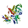
Mass: 18.015 Da / Num. of mol.: 19 / Source method: isolated from a natural source / Formula: H2O

-
Experimental details

-
Experiment

ExperimentMethod: X-RAY DIFFRACTION / Number of used crystals: 1

-
Sample preparation

CrystalDensity Matthews: 2.78 Å3/Da / Density % sol: 55.49 %
Crystal growTemperature: 277 K / Method: vapor diffusion, hanging drop / pH: 7
Details: PEG 800, DTT, HEPES, pH 7.0, VAPOR DIFFUSION, HANGING DROP, temperature 277K

-
Data collection

DiffractionMean temperature: 100 K
Diffraction sourceSource: SYNCHROTRON / Site: CHESS / Beamline: F1 / Wavelength: 0.978 Å
RadiationProtocol: SINGLE WAVELENGTH / Monochromatic (M) / Laue (L): M / Scattering type: x-ray
Radiation wavelengthWavelength: 0.978 Å / Relative weight: 1
ReflectionResolution: 2.8→40 Å / Num. obs: 17214 / % possible obs: 94.8 % / Observed criterion σ(F): 2 / Observed criterion σ(I): 2 / Redundancy: 3.8 % / Rmerge(I) obs: 0.099 / Rsym value: 0.099 / Net I/σ(I): 5.1
Reflection shellResolution: 2.8→2.95 Å / Redundancy: 3.8 % / Rmerge(I) obs: 0.315 / Mean I/σ(I) obs: 2 / Num. unique all: 2210 / Rsym value: 0.315 / % possible all: 89.9

-
Processing

Software
NameVersionClassification
DENZOdata reduction
SCALAdata scaling
AMoREphasing
CNSrefinement
CCP4(SCALA)data scaling
RefinementMethod to determine structure: MOLECULAR REPLACEMENT / Resolution: 2.8→20 Å / σ(F): 0 / Stereochemistry target values: Engh & Huber
RfactorNum. reflectionSelection details
Rfree0.296 1375 10% of observed reflections
Rwork0.233 --
all0.25 --
obs0.241 16788 -
Refinement stepCycle: LAST / Resolution: 2.8→20 Å
ProteinNucleic acidLigandSolventTotal
Num. atoms4043 0 0 19 4062

+
About Yorodumi

-
News

-
Feb 9, 2022. New format data for meta-information of EMDB entries

New format data for meta-information of EMDB entries

  • Version 3 of the EMDB header file is now the official format.
  • The previous official version 1.9 will be removed from the archive.

Related info.:EMDB header

External links:wwPDB to switch to version 3 of the EMDB data model

-
Aug 12, 2020. Covid-19 info

Covid-19 info

URL: https://pdbjlvh1.pdbj.org/emnavi/covid19.php

New page: Covid-19 featured information page in EM Navigator.

Related info.:Covid-19 info / Mar 5, 2020. Novel coronavirus structure data

+
Mar 5, 2020. Novel coronavirus structure data

Novel coronavirus structure data

Related info.:Yorodumi Speices / Aug 12, 2020. Covid-19 info

External links:COVID-19 featured content - PDBj / Molecule of the Month (242):Coronavirus Proteases

+
Jan 31, 2019. EMDB accession codes are about to change! (news from PDBe EMDB page)

EMDB accession codes are about to change! (news from PDBe EMDB page)

  • The allocation of 4 digits for EMDB accession codes will soon come to an end. Whilst these codes will remain in use, new EMDB accession codes will include an additional digit and will expand incrementally as the available range of codes is exhausted. The current 4-digit format prefixed with “EMD-” (i.e. EMD-XXXX) will advance to a 5-digit format (i.e. EMD-XXXXX), and so on. It is currently estimated that the 4-digit codes will be depleted around Spring 2019, at which point the 5-digit format will come into force.
  • The EM Navigator/Yorodumi systems omit the EMD- prefix.

Related info.:Q: What is EMD? / ID/Accession-code notation in Yorodumi/EM Navigator

External links:EMDB Accession Codes are Changing Soon! / Contact to PDBj

+
Jul 12, 2017. Major update of PDB

Major update of PDB

  • wwPDB released updated PDB data conforming to the new PDBx/mmCIF dictionary.
  • This is a major update changing the version number from 4 to 5, and with Remediation, in which all the entries are updated.
  • In this update, many items about electron microscopy experimental information are reorganized (e.g. em_software).
  • Now, EM Navigator and Yorodumi are based on the updated data.

External links:wwPDB Remediation / Enriched Model Files Conforming to OneDep Data Standards Now Available in the PDB FTP Archive

-
Yorodumi

Thousand views of thousand structures

  • Yorodumi is a browser for structure data from EMDB, PDB, SASBDB, etc.
  • This page is also the successor to EM Navigator detail page, and also detail information page/front-end page for Omokage search.
  • The word "yorodu" (or yorozu) is an old Japanese word meaning "ten thousand". "mi" (miru) is to see.

Related info.:EMDB / PDB / SASBDB / Comparison of 3 databanks / Yorodumi Search / Aug 31, 2016. New EM Navigator & Yorodumi / Yorodumi Papers / Jmol/JSmol / Function and homology information / Changes in new EM Navigator and Yorodumi

Read more