 Movie
Movie Controller


+ Open data

- Basic information
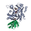
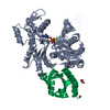
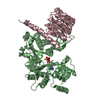
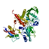
| Entry | Database: PDB / ID: 2btf | ||||||
|---|---|---|---|---|---|---|---|
| Title | THE STRUCTURE OF CRYSTALLINE PROFILIN-BETA-ACTIN | ||||||
 Components Components |
| ||||||
 Keywords Keywords | ACETYLATION AND ACTIN-BINDING | ||||||
| Function / homology |  Function and homology information Function and homology informationPCP/CE pathway / Cell-extracellular matrix interactions / Formation of the canonical BAF (cBAF) complex / Formation of the polybromo-BAF (pBAF) complex / Formation of the embryonic stem cell BAF (esBAF) complex / Formation of the non-canonical BAF (ncBAF) complex / Formation of neuronal progenitor and neuronal BAF (npBAF and nBAF) / Adherens junctions interactions / Regulation of CDH1 Function / Formation of the dystrophin-glycoprotein complex (DGC) ...PCP/CE pathway / Cell-extracellular matrix interactions / Formation of the canonical BAF (cBAF) complex / Formation of the polybromo-BAF (pBAF) complex / Formation of the embryonic stem cell BAF (esBAF) complex / Formation of the non-canonical BAF (ncBAF) complex / Formation of neuronal progenitor and neuronal BAF (npBAF and nBAF) / Adherens junctions interactions / Regulation of CDH1 Function / Formation of the dystrophin-glycoprotein complex (DGC) / B-WICH complex positively regulates rRNA expression / Gap junction degradation / Formation of annular gap junctions / RHOF GTPase cycle / EPHB-mediated forward signaling / MAP2K and MAPK activation / Regulation of actin dynamics for phagocytic cup formation / RHO GTPases Activate WASPs and WAVEs / RHO GTPases activate IQGAPs / RHO GTPases Activate Formins / DNA Damage Recognition in GG-NER / UCH proteinases / VEGFA-VEGFR2 Pathway / positive regulation of actin filament bundle assembly / regulation of actin filament polymerization / cellular response to cytochalasin B / Clathrin-mediated endocytosis / regulation of transepithelial transport / morphogenesis of a polarized epithelium / structural constituent of postsynaptic actin cytoskeleton / protein localization to adherens junction / dense body / Tat protein binding / postsynaptic actin cytoskeleton / adherens junction assembly / apical protein localization / tight junction / apical junction complex / regulation of norepinephrine uptake / transporter regulator activity / NuA4 histone acetyltransferase complex / cortical cytoskeleton / establishment or maintenance of cell polarity / nitric-oxide synthase binding / brush border / regulation of synaptic vesicle endocytosis / kinesin binding / regulation of protein localization to plasma membrane / positive regulation of double-strand break repair via homologous recombination / axonogenesis / calyx of Held / nitric-oxide synthase regulator activity / actin filament / adherens junction / cell motility / Hydrolases; Acting on acid anhydrides; Acting on acid anhydrides to facilitate cellular and subcellular movement / Schaffer collateral - CA1 synapse / cytoplasmic ribonucleoprotein granule / nucleosome / lamellipodium / actin cytoskeleton / actin binding / actin cytoskeleton organization / cytoskeleton / regulation of cell cycle / ribonucleoprotein complex / axon / focal adhesion / synapse / protein kinase binding / glutamatergic synapse / ATP hydrolysis activity / protein-containing complex / ATP binding / membrane / identical protein binding / nucleus / plasma membrane / cytoplasm / cytosol Similarity search - Function | ||||||
| Biological species |  | ||||||
| Method |  X-RAY DIFFRACTION / Resolution: 2.55 Å X-RAY DIFFRACTION / Resolution: 2.55 Å | ||||||
 Authors Authors | Schutt, C.E. / Myslik, J.C. / Rozycki, M.D. / Goonesekere, N.C.W. | ||||||
 Citation Citation | Journal: Nature / Year: 1993 Title: The structure of crystalline profilin-beta-actin. Authors: Schutt, C.E. / Myslik, J.C. / Rozycki, M.D. / Goonesekere, N.C. / Lindberg, U. #1: Journal: To be PublishedTitle: Structural Aspects of Actin Binding Proteins Current Opinion in Cell Biology Authors: Rozycki, M.D. / Myslik, J.C. / Schutt, C.E. / Lindberg, U. #2: Journal: FEBS Lett. / Year: 1993Title: Mutagenesis of Human Profilin Locates its Poly(L-Proline)-Binding Site to a Hydrophobic Patch of Aromatic Amino Acids Authors: Bjorkegren, C. / Rozycki, M. / Schutt, C.E. / Lindberg, U. / Karlsson, R. #3: Journal: J.Mol.Biol. / Year: 1989Title: Molecular Packing in Profilin-Actin Crystals and its Implications Authors: Schutt, C.E. / Lindberg, U. / Myslik, J. / Strauss, N. | ||||||
| History |
| ||||||
| Remark 650 | HELIX RESIDUES ARE INCLUDED AT THE BEGINNING OF HELICES IF THEY PARTICIPATE IN HYDROGEN BONDING AND ...HELIX RESIDUES ARE INCLUDED AT THE BEGINNING OF HELICES IF THEY PARTICIPATE IN HYDROGEN BONDING AND ARE "PARTIALLY" HELICAL, EVEN IF THE ENTIRE RESIDUE DOES NOT FIT HELICAL CRITERIA. | ||||||
| Remark 700 | SHEET PROFILIN IS DESCRIBED IN JRNL REFERENCE AS HAVING A SIX-MEMBERED BETA-SHEET WHICH SHARES A ...SHEET PROFILIN IS DESCRIBED IN JRNL REFERENCE AS HAVING A SIX-MEMBERED BETA-SHEET WHICH SHARES A STRAND WITH A SECOND TWO-MEMBERED SHEET. TO CONFORM WITH PROTEIN DATA BANK GUIDELINES, THESE SHEETS ARE COMBINED INTO A SINGLE SEVEN-MEMBERED SHEET (SP1) IN THIS ENTRY. STRAND NUMBERING ALSO DIFFERS FROM THE JRNL REFERENCE. SECONDARY STRUCTURE ASSIGNMENTS FOR BETA-ACTIN ARE SIMILAR TO THOSE USED FOR ALPHA-ACTIN (PROTEIN DATA BANK ENTRY 1ATN) WITH MINOR CHANGES. HOWEVER, THE NOMENCLATURE USED HEREIN DIFFERS FROM THAT USED IN THE ABOVE ENTRY. ALSO, STRAND A4B DESCRIBED IN THAT ENTRY IS NOT INCLUDED HERE BECAUSE THE "ALTERNATIVE" STRAND TO ONE IN STRAND A4A (CORRESPONDING TO SA4 IN THIS ENTRY) IS ONLY TWO RESIDUES IN LENGTH. IN CONTRAST, SHEET SA3 IS INCLUDED BECAUSE IT CONTAINS TWO STRANDS AND A WELL-DEFINED TYPE II TURN, EVEN THOUGH THE TWO STRANDS CONTAIN ONLY TWO RESIDUES EACH. RESIDUE P 31 IS A BETA-BULGE. RESIDUE P 73 IS A BETA-BULGE, REDIRECTING STRAND 6. |
- Structure visualization
| Structure viewer | Molecule: MolmilJmol/JSmol |
|---|
- Downloads & links
-Download
| PDBx/mmCIF format | 2btf.cif.gz | 113.6 KB | Display | PDBx/mmCIF format |
|---|---|---|---|---|
| PDB format | pdb2btf.ent.gz | 86.3 KB | Display | PDB format |
| PDBx/mmJSON format | 2btf.json.gz | Tree view | PDBx/mmJSON format | |
| Others |  Other downloads Other downloads |
-Validation report
| Arichive directory | https://data.pdbj.org/pub/pdb/validation_reports/bt/2btfftp://data.pdbj.org/pub/pdb/validation_reports/bt/2btf | HTTPS FTP |
|---|
-Related structure data
| Similar structure data |
|---|


-Links
 PDBj
PDBj


- Assembly
| Deposited unit | 
| ||||||||
|---|---|---|---|---|---|---|---|---|---|
| 1 |
| ||||||||
| Unit cell |
| ||||||||
| Atom site foot note | 1: RESIDUE ACE A IS THE N-ACETYL MOIETY OF ACTIN. / 2: RESIDUE A 73 IS 3-METHYL HISTIDYL. / 3: RESIDUE ACE P IS THE N-ACETYL MOIETY OF PROFILIN. / 4: RESIDUE P 31 IS A BETA-BULGE. / 5: RESIDUE P 73 IS A BETA-BULGE, REDIRECTING STRAND 6. |
-Components
| #1: Protein | Mass: 41690.520 Da / Num. of mol.: 1 Source method: isolated from a genetically manipulated source Source: (gene. exp.) |
|---|---|
| #2: Protein | Mass: 14968.185 Da / Num. of mol.: 1 Source method: isolated from a genetically manipulated source Source: (gene. exp.) |
| #3: Chemical | ChemComp-SR /   Mass: 87.620 Da / Num. of mol.: 1 / Source method: obtained synthetically / Formula: Sr Mass: 87.620 Da / Num. of mol.: 1 / Source method: obtained synthetically / Formula: Sr |
| #4: Chemical | ChemComp-ATP /   Mass: 507.181 Da / Num. of mol.: 1 / Source method: obtained synthetically / Formula: C10H16N5O13P3 / Comment: ATP, energy-carrying molecule*YM Mass: 507.181 Da / Num. of mol.: 1 / Source method: obtained synthetically / Formula: C10H16N5O13P3 / Comment: ATP, energy-carrying molecule*YM |
| Has protein modification | Y |
-Experimental details
-Experiment
| Experiment | Method: X-RAY DIFFRACTION |
|---|
- Sample preparation
| Crystal | Density Matthews: 2.11 Å3/Da / Density % sol: 41.6 % | ||||||||||||||||||||||||||||||
|---|---|---|---|---|---|---|---|---|---|---|---|---|---|---|---|---|---|---|---|---|---|---|---|---|---|---|---|---|---|---|---|
| Crystal grow | *PLUS Temperature: 4 ℃ / Method: vapor diffusion, hanging dropDetails: referred to 'Segura, M. & Lindberg, U.', (1984) J.Biol.Chem., 259, 3949-3954 | ||||||||||||||||||||||||||||||
| Components of the solutions | *PLUS
|
-Data collection
| Radiation | Scattering type: x-ray |
|---|---|
| Radiation wavelength | Relative weight: 1 |
| Reflection | *PLUS Highest resolution: 2.55 Å / Num. obs: 16158 / % possible obs: 98.8 % / Observed criterion σ(I): 0 / Rmerge(I) obs: 0.065 |
- Processing
| Software |
| ||||||||||||||||||||||||||||||||||||||||||||||||||||||||||||
|---|---|---|---|---|---|---|---|---|---|---|---|---|---|---|---|---|---|---|---|---|---|---|---|---|---|---|---|---|---|---|---|---|---|---|---|---|---|---|---|---|---|---|---|---|---|---|---|---|---|---|---|---|---|---|---|---|---|---|---|---|---|
| Refinement | Rfactor Rwork: 0.199 / Rfactor obs: 0.199 / Highest resolution: 2.55 Å | ||||||||||||||||||||||||||||||||||||||||||||||||||||||||||||
| Refinement step | Cycle: LAST / Highest resolution: 2.55 Å
| ||||||||||||||||||||||||||||||||||||||||||||||||||||||||||||
| Refine LS restraints |
| ||||||||||||||||||||||||||||||||||||||||||||||||||||||||||||
| Refinement | *PLUS Lowest resolution: 8 Å / σ(F): 3 / Rfactor obs: 0.199 / Rfactor Rwork: 0.199 | ||||||||||||||||||||||||||||||||||||||||||||||||||||||||||||
| Solvent computation | *PLUS | ||||||||||||||||||||||||||||||||||||||||||||||||||||||||||||
| Displacement parameters | *PLUS Biso mean: 19.9 Å2 | ||||||||||||||||||||||||||||||||||||||||||||||||||||||||||||
| Refine LS restraints | *PLUS Type: x_angle_d / Dev ideal: 3.8 | ||||||||||||||||||||||||||||||||||||||||||||||||||||||||||||
| LS refinement shell | *PLUS Highest resolution: 2.55 Å / Lowest resolution: 2.66 Å / Total num. of bins used: 8 / Num. reflection obs: 1539 / Rfactor obs: 0.2961 |
