 Movie
Movie Controller

[English] 日本語
 Yorodumi
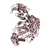
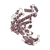
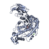
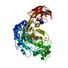
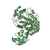
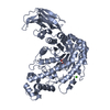
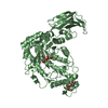
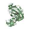
- PDB-1j0y: Beta-amylase from Bacillus cereus var. mycoides in complex with g... -
+ Open data

- Basic information
| Entry | Database: PDB / ID: 1j0y | ||||||
|---|---|---|---|---|---|---|---|
| Title | Beta-amylase from Bacillus cereus var. mycoides in complex with glucose | ||||||
 Components Components | Beta-amylase | ||||||
 Keywords Keywords | HYDROLASE / BETA-AMYLASE / RAW-STARCH BINDING DOMAIN | ||||||
| Function / homology |  Function and homology information Function and homology informationbeta-amylase / beta-amylase activity / starch binding / polysaccharide catabolic process / metal ion binding Similarity search - Function | ||||||
| Biological species |  | ||||||
| Method |  X-RAY DIFFRACTION / FOURIER SYNTHESIS / Resolution: 2.1 Å X-RAY DIFFRACTION / FOURIER SYNTHESIS / Resolution: 2.1 Å | ||||||
 Authors Authors | Oyama, T. / Miyake, H. / Kusunoki, M. / Nitta, Y. | ||||||
 Citation Citation | Journal: J.BIOCHEM.(TOKYO) / Year: 2003 Title: Crystal Structures of beta-Amylase from Bacillus cereus var. mycoides in Complexes with Substrate Analogs and Affinity-Labeling Reagents Authors: Oyama, T. / Miyake, H. / Kusunoki, M. / Nitta, Y. #1: Journal: J.BIOCHEM.(TOKYO) / Year: 1999Title: Crystal Structure of beta-Amylase from Bacillus cereus var. mycoides at 2.2 A resolution Authors: Oyama, T. / Kusunoki, M. / Kishimoto, Y. / Takasaki, Y. / Nitta, Y. #2: Journal: BIOSCI.BIOTECHNOL.BIOCHEM. / Year: 1996Title: Kinetic Study of Active Site Structure of beta-Amylase from Bacillus cereus var. mycoides Authors: Nitta, Y. / Shirakawa, M. / Takasaki, Y. | ||||||
| History |
|
- Structure visualization
| Structure viewer | Molecule: MolmilJmol/JSmol |
|---|
- Downloads & links
-Download
| PDBx/mmCIF format | 1j0y.cif.gz | 416.8 KB | Display | PDBx/mmCIF format |
|---|---|---|---|---|
| PDB format | pdb1j0y.ent.gz | 342.5 KB | Display | PDB format |
| PDBx/mmJSON format | 1j0y.json.gz | Tree view | PDBx/mmJSON format | |
| Others |  Other downloads Other downloads |
-Validation report
| Arichive directory | https://data.pdbj.org/pub/pdb/validation_reports/j0/1j0yftp://data.pdbj.org/pub/pdb/validation_reports/j0/1j0y | HTTPS FTP |
|---|
-Related structure data
| Related structure data | 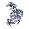 1j0zC  1j10C  1j11C  1j12C 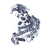 5bcaS S: Starting model for refinement C: citing same article ( |
|---|---|
| Similar structure data |

-Links
 PDBj
PDBj


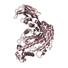
- Assembly
| Deposited unit | 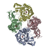
| ||||||||
|---|---|---|---|---|---|---|---|---|---|
| 1 | 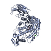
| ||||||||
| 2 | 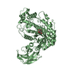
| ||||||||
| 3 | 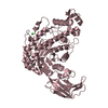
| ||||||||
| 4 | 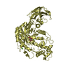
| ||||||||
| Unit cell |
|
-Components
| #1: Protein | Mass: 58358.523 Da / Num. of mol.: 4 / Source method: isolated from a natural source / Source: (natural) #2: Sugar | ChemComp-BGC /   Type: D-saccharide, beta linking / Mass: 180.156 Da / Num. of mol.: 4 Type: D-saccharide, beta linking / Mass: 180.156 Da / Num. of mol.: 4Source method: isolated from a genetically manipulated source Formula: C6H12O6 #3: Chemical | ChemComp-CA /   Mass: 40.078 Da / Num. of mol.: 4 / Source method: obtained synthetically / Formula: Ca Mass: 40.078 Da / Num. of mol.: 4 / Source method: obtained synthetically / Formula: Ca#4: Water | ChemComp-HOH / |  Mass: 18.015 Da / Num. of mol.: 635 / Source method: isolated from a natural source / Formula: H2O Mass: 18.015 Da / Num. of mol.: 635 / Source method: isolated from a natural source / Formula: H2OHas protein modification | Y | |
|---|
-Experimental details
-Experiment
| Experiment | Method: X-RAY DIFFRACTION / Number of used crystals: 1 |
|---|
- Sample preparation
| Crystal | Density Matthews: 2.94 Å3/Da / Density % sol: 57.83 % | |||||||||||||||||||||||||
|---|---|---|---|---|---|---|---|---|---|---|---|---|---|---|---|---|---|---|---|---|---|---|---|---|---|---|
| Crystal grow | Temperature: 293 K / Method: vapor diffusion, hanging drop / pH: 9 Details: PEG 6000, pH 9.0, VAPOR DIFFUSION, HANGING DROP, temperature 293K | |||||||||||||||||||||||||
| Crystal grow | *PLUS Temperature: 293 K / Method: vapor diffusion, hanging drop / Details: Oyama, T., (1998) Protein Pept.Lett., 5, 349. | |||||||||||||||||||||||||
| Components of the solutions | *PLUS
|
-Data collection
| Diffraction | Mean temperature: 293 K |
|---|---|
| Diffraction source | Source: ROTATING ANODE / Type: RIGAKU / Wavelength: 1.5418 Å |
| Detector | Type: RIGAKU RAXIS IV / Detector: IMAGE PLATE / Date: Oct 1, 1998 / Details: mirrors |
| Radiation | Monochromator: YALE MIRRORS / Protocol: SINGLE WAVELENGTH / Monochromatic (M) / Laue (L): M / Scattering type: x-ray |
| Radiation wavelength | Wavelength: 1.5418 Å / Relative weight: 1 |
| Reflection | Resolution: 2→39.5 Å / Num. obs: 135775 / % possible obs: 72.8 % / Observed criterion σ(I): 0 / Rmerge(I) obs: 0.062 |
| Reflection shell | Resolution: 2→2.09 Å / Rmerge(I) obs: 0.214 / % possible all: 53.1 |
| Reflection | *PLUS Highest resolution: 2 Å / Redundancy: 1.8 % / Num. measured all: 243229 |
| Reflection shell | *PLUS % possible obs: 53.1 % / Redundancy: 1.5 % / Num. unique obs: 12350 / Mean I/σ(I) obs: 3.1 |
- Processing
| Software |
| ||||||||||||||||||||
|---|---|---|---|---|---|---|---|---|---|---|---|---|---|---|---|---|---|---|---|---|---|
| Refinement | Method to determine structure: FOURIER SYNTHESIS Starting model: PDB ENTRY 5BCA Resolution: 2.1→8 Å / Isotropic thermal model: Isotropic / Cross valid method: THROUGHOUT / σ(F): 2 / Stereochemistry target values: Engh & Huber
| ||||||||||||||||||||
| Refinement step | Cycle: LAST / Resolution: 2.1→8 Å
| ||||||||||||||||||||
| Refine LS restraints |
| ||||||||||||||||||||
| Refinement | *PLUS % reflection Rfree: 5 % | ||||||||||||||||||||
| Solvent computation | *PLUS | ||||||||||||||||||||
| Displacement parameters | *PLUS | ||||||||||||||||||||
| Refine LS restraints | *PLUS
|
