 Movie
Movie Controller

[English] 日本語
 Yorodumi
- PDB-2a6d: Crystal structure analysis of the anti-arsonate germline antibody... -
+ Open data

- Basic information
| Entry | Database: PDB / ID: 2a6d | ||||||
|---|---|---|---|---|---|---|---|
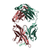
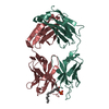
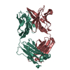
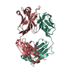
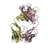
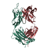
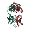
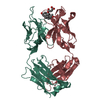
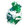
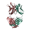
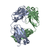
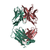
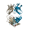
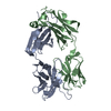
| Title | Crystal structure analysis of the anti-arsonate germline antibody 36-65 in complex with a phage display derived dodecapeptide RLLIADPPSPRE | ||||||
 Components Components |
| ||||||
 Keywords Keywords | IMMUNE SYSTEM / Germline / FAB / ANTIBODY | ||||||
| Function / homology | Immunoglobulins / Immunoglobulin-like / Sandwich / Mainly Beta Function and homology information Function and homology information | ||||||
| Biological species |  | ||||||
| Method |  X-RAY DIFFRACTION / MOLECULAR REPLACEMENT / Resolution: 2.9 Å X-RAY DIFFRACTION / MOLECULAR REPLACEMENT / Resolution: 2.9 Å | ||||||
 Authors Authors | Sethi, D.K. / Agarwal, A. / Manivel, V. / Rao, K.V. / Salunke, D.M. | ||||||
 Citation Citation | Journal: Immunity / Year: 2006 Title: Differential epitope positioning within the germline antibody paratope enhances promiscuity in the primary immune response. Authors: Sethi, D.K. / Agarwal, A. / Manivel, V. / Rao, K.V. / Salunke, D.M. | ||||||
| History |
| ||||||
| Remark 999 | SEQUENCE THE SEQUENCE IS NOT DEPOSITED IN ANY SEQUENCE DATABASE EXCEPT RESIDUES 1 TO 120 OF CHAINS ...SEQUENCE THE SEQUENCE IS NOT DEPOSITED IN ANY SEQUENCE DATABASE EXCEPT RESIDUES 1 TO 120 OF CHAINS B,H WHICH CORRESPOND TO DEPOSITED SEQUENCE OF THE HEAVY CHAIN VARIABLE REGION OF ANTI-ARSONATE ANTIBODY 36-65 |
- Structure visualization
| Structure viewer | Molecule: MolmilJmol/JSmol |
|---|
- Downloads & links
-Download
| PDBx/mmCIF format | 2a6d.cif.gz | 176.3 KB | Display | PDBx/mmCIF format |
|---|---|---|---|---|
| PDB format | pdb2a6d.ent.gz | 140.6 KB | Display | PDB format |
| PDBx/mmJSON format | 2a6d.json.gz | Tree view | PDBx/mmJSON format | |
| Others |  Other downloads Other downloads |
-Validation report
| Arichive directory | https://data.pdbj.org/pub/pdb/validation_reports/a6/2a6dftp://data.pdbj.org/pub/pdb/validation_reports/a6/2a6d | HTTPS FTP |
|---|
-Related structure data
| Related structure data | 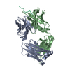 2a6iC 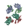 2a6jC 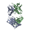 2a6kC  6fabS S: Starting model for refinement C: citing same article ( |
|---|---|
| Similar structure data |


-Links
 PDBj
PDBj

- Assembly
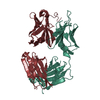
| Deposited unit | 
| ||||||||
|---|---|---|---|---|---|---|---|---|---|
| 1 | 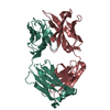
| ||||||||
| 2 | 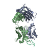
| ||||||||
| Unit cell |
|
-Components
| #1: Antibody | Mass: 23707.049 Da / Num. of mol.: 2 / Fragment: Fab / Source method: isolated from a natural source / Details: Mouse monoclonal / Source: (natural) #2: Antibody | Mass: 23942.768 Da / Num. of mol.: 2 / Source method: isolated from a natural source / Details: Mouse monoclonal / Source: (natural) #3: Protein/peptide | | Mass: 1365.578 Da / Num. of mol.: 1 / Source method: obtained synthetically Details: chemically synthesized (Phage display derived peptide) #4: Water | ChemComp-HOH / |  Mass: 18.015 Da / Num. of mol.: 78 / Source method: isolated from a natural source / Formula: H2O Mass: 18.015 Da / Num. of mol.: 78 / Source method: isolated from a natural source / Formula: H2OHas protein modification | Y | |
|---|
-Experimental details
-Experiment
| Experiment | Method: X-RAY DIFFRACTION / Number of used crystals: 1 |
|---|
- Sample preparation
| Crystal | Density Matthews: 2.7 Å3/Da / Density % sol: 53.8 % |
|---|---|
| Crystal grow | Temperature: 298 K / Method: vapor diffusion, hanging drop / pH: 6.7 Details: 18% PEG 8000, cacodylate, 0.05% azide, pH 6.7, VAPOR DIFFUSION, HANGING DROP, temperature 298.0K |
-Data collection
| Diffraction | Mean temperature: 120 K |
|---|---|
| Diffraction source | Source: ROTATING ANODE / Type: RIGAKU RU200 / Wavelength: 1.5418 Å |
| Detector | Type: MARRESEARCH / Detector: IMAGE PLATE / Date: Jan 1, 2003 / Details: monochromator |
| Radiation | Protocol: SINGLE WAVELENGTH / Monochromatic (M) / Laue (L): M / Scattering type: x-ray |
| Radiation wavelength | Wavelength: 1.5418 Å / Relative weight: 1 |
| Reflection | Resolution: 2.9→100 Å / Num. all: 23421 / Num. obs: 23421 / % possible obs: 83 % / Observed criterion σ(F): 0 / Observed criterion σ(I): 0 / Redundancy: 4.2 % / Rmerge(I) obs: 0.13 / Net I/σ(I): 6.87 |
- Processing
| Software |
| |||||||||||||||||||||||||
|---|---|---|---|---|---|---|---|---|---|---|---|---|---|---|---|---|---|---|---|---|---|---|---|---|---|---|
| Refinement | Method to determine structure: MOLECULAR REPLACEMENT Starting model: 6FAB Resolution: 2.9→100 Å / Cross valid method: THROUGHOUT / σ(F): 0 / Stereochemistry target values: Engh & Huber
| |||||||||||||||||||||||||
| Refinement step | Cycle: LAST / Resolution: 2.9→100 Å
| |||||||||||||||||||||||||
| Refine LS restraints |
|
