 Movie
Movie Controller

[English] 日本語
 Yorodumi
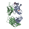
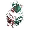
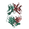
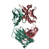
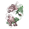
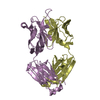
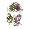
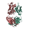
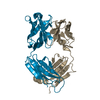
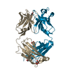
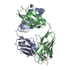
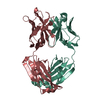
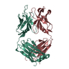
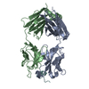
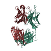
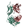
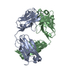
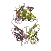
- PDB-2h1p: THE THREE-DIMENSIONAL STRUCTURES OF A POLYSACCHARIDE BINDING ANTI... -
+ Open data

- Basic information
| Entry | Database: PDB / ID: 2h1p | ||||||
|---|---|---|---|---|---|---|---|
| Title | THE THREE-DIMENSIONAL STRUCTURES OF A POLYSACCHARIDE BINDING ANTIBODY TO CRYPTOCOCCUS NEOFORMANS AND ITS COMPLEX WITH A PEPTIDE FROM A PHAGE DISPLAY LIBRARY: IMPLICATIONS FOR THE IDENTIFICATION OF PEPTIDE MIMOTOPES | ||||||
 Components Components |
| ||||||
 Keywords Keywords | COMPLEX (ANTIBODY/PEPTIDE) / ANTIBODY STRUCTURE / CRYPTOCOCCUS / PHAGE LIBRARY / POLYSACCHARIDE / COMPLEX (ANTIBODY-PEPTIDE) / COMPLEX (ANTIBODY-PEPTIDE) complex | ||||||
| Function / homology | Immunoglobulins / Immunoglobulin-like / Sandwich / Mainly Beta / : / :  Function and homology information Function and homology information | ||||||
| Biological species |  | ||||||
| Method |  X-RAY DIFFRACTION / MOLECULAR REPLACEMENT / Resolution: 2.4 Å X-RAY DIFFRACTION / MOLECULAR REPLACEMENT / Resolution: 2.4 Å | ||||||
 Authors Authors | Young, A.C.M. / Valadon, P. / Casadevall, A. / Scharff, M.D. / Sacchettini, J.C. | ||||||
 Citation Citation | Journal: J.Mol.Biol. / Year: 1997 Title: The three-dimensional structures of a polysaccharide binding antibody to Cryptococcus neoformans and its complex with a peptide from a phage display library: implications for the ...Title: The three-dimensional structures of a polysaccharide binding antibody to Cryptococcus neoformans and its complex with a peptide from a phage display library: implications for the identification of peptide mimotopes. Authors: Young, A.C. / Valadon, P. / Casadevall, A. / Scharff, M.D. / Sacchettini, J.C. #1: Journal: J.Mol.Biol. / Year: 1996Title: Peptide Libraries Define the Fine Specificity of Anti-Polysaccharide Antibodies to Cryptococcus Neoformans Authors: Valadon, P. / Nussbaum, G. / Boyd, L.F. / Margulies, D.H. / Scharff, M.D. | ||||||
| History |
|
- Structure visualization
| Structure viewer | Molecule: MolmilJmol/JSmol |
|---|
- Downloads & links
-Download
| PDBx/mmCIF format | 2h1p.cif.gz | 100.6 KB | Display | PDBx/mmCIF format |
|---|---|---|---|---|
| PDB format | pdb2h1p.ent.gz | 75.8 KB | Display | PDB format |
| PDBx/mmJSON format | 2h1p.json.gz | Tree view | PDBx/mmJSON format | |
| Others |  Other downloads Other downloads |
-Validation report
| Arichive directory | https://data.pdbj.org/pub/pdb/validation_reports/h1/2h1pftp://data.pdbj.org/pub/pdb/validation_reports/h1/2h1p | HTTPS FTP |
|---|
-Related structure data
| Related structure data |  1flrS S: Starting model for refinement |
|---|---|
| Similar structure data |


-Links
 PDBj
PDBj

- Assembly
| Deposited unit | 
| ||||||||
|---|---|---|---|---|---|---|---|---|---|
| 1 |
| ||||||||
| Unit cell |
|
-Components
| #1: Antibody | Mass: 24125.727 Da / Num. of mol.: 1 / Fragment: FAB / Source method: isolated from a natural source Details: FAB PART ISOLATED AFTER PAPAIN DIGESTION OF THE PARENT IGG1/K MOLECULE Source: (natural) |
|---|---|
| #2: Antibody | Mass: 23687.664 Da / Num. of mol.: 1 / Fragment: FAB / Source method: isolated from a natural source Details: FAB PART ISOLATED AFTER PAPAIN DIGESTION OF THE PARENT IGG1/K MOLECULE Source: (natural) |
| #3: Protein/peptide | Mass: 1351.570 Da / Num. of mol.: 1 Source method: isolated from a genetically manipulated source |
| #4: Water | ChemComp-HOH /  Mass: 18.015 Da / Num. of mol.: 62 / Source method: isolated from a natural source / Formula: H2O Mass: 18.015 Da / Num. of mol.: 62 / Source method: isolated from a natural source / Formula: H2O |
| Has protein modification | Y |
-Experimental details
-Experiment
| Experiment | Method: X-RAY DIFFRACTION / Number of used crystals: 2 |
|---|
- Sample preparation
| Crystal | Density Matthews: 3.28 Å3/Da / Density % sol: 62.2 % | ||||||||||||||||||||
|---|---|---|---|---|---|---|---|---|---|---|---|---|---|---|---|---|---|---|---|---|---|
| Crystal grow | pH: 8.5 / Details: 18% PEG 8K, PH 8.5, 1% GLYCEROL | ||||||||||||||||||||
| Crystal grow | *PLUS Method: vapor diffusion, hanging drop | ||||||||||||||||||||
| Components of the solutions | *PLUS
|
-Data collection
| Diffraction | Mean temperature: 290 K |
|---|---|
| Diffraction source | Source: ROTATING ANODE / Type: RIGAKU RUH2R / Wavelength: 1.5418 |
| Detector | Type: SIEMENS / Detector: AREA DETECTOR / Date: Sep 1, 1995 / Details: COLLIMATOR |
| Radiation | Monochromator: CU K ALPHA / Monochromatic (M) / Laue (L): M / Scattering type: x-ray |
| Radiation wavelength | Wavelength: 1.5418 Å / Relative weight: 1 |
| Reflection | Resolution: 2.4→20 Å / Num. obs: 19345 / % possible obs: 90 % / Redundancy: 2.4 % / Rmerge(I) obs: 0.065 / Net I/σ(I): 15.3 |
| Reflection shell | Resolution: 2.4→2.5 Å / % possible all: 79 |
| Reflection shell | *PLUS % possible obs: 79 % |
- Processing
| Software |
| ||||||||||||||||||||||||||||||
|---|---|---|---|---|---|---|---|---|---|---|---|---|---|---|---|---|---|---|---|---|---|---|---|---|---|---|---|---|---|---|---|
| Refinement | Method to determine structure: MOLECULAR REPLACEMENT Starting model: PDB ENTRY 1FLR (FAB 4-4-20) Resolution: 2.4→20 Å / Isotropic thermal model: TNT / σ(F): 2 / Stereochemistry target values: TNT
| ||||||||||||||||||||||||||||||
| Solvent computation | Solvent model: TNT | ||||||||||||||||||||||||||||||
| Refinement step | Cycle: LAST / Resolution: 2.4→20 Å
| ||||||||||||||||||||||||||||||
| Refine LS restraints |
| ||||||||||||||||||||||||||||||
| Software | *PLUS Name: TNT / Classification: refinement | ||||||||||||||||||||||||||||||
| Refinement | *PLUS Rfactor obs: 0.186 | ||||||||||||||||||||||||||||||
| Solvent computation | *PLUS | ||||||||||||||||||||||||||||||
| Displacement parameters | *PLUS |
