 Movie
Movie Controller


+ Open data

- Basic information
| Entry | Database: PDB / ID: 1zf2 | ||||||||||||||||||
|---|---|---|---|---|---|---|---|---|---|---|---|---|---|---|---|---|---|---|---|
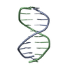
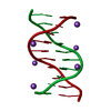
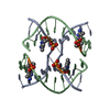
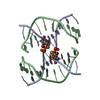
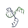
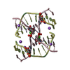
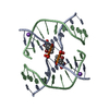
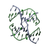
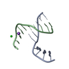
| Title | Four-stranded DNA Holliday Junction (CCC) | ||||||||||||||||||
 Components Components | 5'-D(* Keywords KeywordsDNA / Crystallographic Screen / DNA Structure / Holliday Junction / Molecular Structure | Function / homology | DNA |  Function and homology information Function and homology informationMethod |  X-RAY DIFFRACTION / SYNCHROTRON / MOLECULAR REPLACEMENT / Resolution: 1.95 Å X-RAY DIFFRACTION / SYNCHROTRON / MOLECULAR REPLACEMENT / Resolution: 1.95 Å  Authors AuthorsHays, F.A. / Teegarden, A.T. / Jones, Z.J.R. / Harms, M. / Raup, D. / Watson, J. / Cavaliere, E. / Ho, P.S. |  CitationJournal: Proc.Natl.Acad.Sci.Usa / Year: 2005 CitationJournal: Proc.Natl.Acad.Sci.Usa / Year: 2005Title: How sequence defines structure: a crystallographic map of DNA structure and conformation. Authors: Hays, F.A. / Teegarden, A. / Jones, Z.J. / Harms, M. / Raup, D. / Watson, J. / Cavaliere, E. / Ho, P.S. History |
|
- Structure visualization
| Structure viewer | Molecule: MolmilJmol/JSmol |
|---|
- Downloads & links
-Download
| PDBx/mmCIF format | 1zf2.cif.gz | 20.1 KB | Display | PDBx/mmCIF format |
|---|---|---|---|---|
| PDB format | pdb1zf2.ent.gz | 13.8 KB | Display | PDB format |
| PDBx/mmJSON format | 1zf2.json.gz | Tree view | PDBx/mmJSON format | |
| Others |  Other downloads Other downloads |
-Validation report
| Arichive directory | https://data.pdbj.org/pub/pdb/validation_reports/zf/1zf2ftp://data.pdbj.org/pub/pdb/validation_reports/zf/1zf2 | HTTPS FTP |
|---|
-Related structure data
| Related structure data | 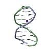 1zewC  1zexC 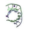 1zeyC 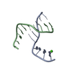 1zezC  1zf0C 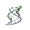 1zf1C  1zf3C 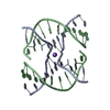 1zf4C 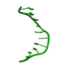 1zf5C 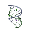 1zf6C 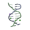 1zf7C 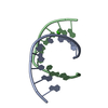 1zf8C 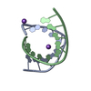 1zf9C 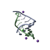 1zfaC 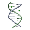 1zfbC 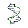 1zfcC  1zfeC 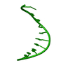 1zffC 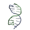 1zfgC  1zfhC 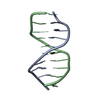 1zfmC C: citing same article ( |
|---|---|
| Similar structure data |


-Links
 PDBj
PDBj

- Assembly
| Deposited unit | 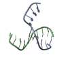
| ||||||||
|---|---|---|---|---|---|---|---|---|---|
| 1 | 
| ||||||||
| Unit cell |
| ||||||||
| Components on special symmetry positions |
|
-Components
| #1: DNA chain | Mass: 3046.980 Da / Num. of mol.: 2 / Source method: obtained synthetically Details: DNA WAS SYNTHESIZED ON AN APPLIED BIOSYSTEMS DNA SYNTHESIZER USING PHOSPHORAMIDITE CHEMISTRY, WITH THE TRITYL-PROTECTING GROUP LEFT INTACT AT THE 5'-TERMINAL NUCLEOTIDE THEN DEPROTECTED BY ...Details: DNA WAS SYNTHESIZED ON AN APPLIED BIOSYSTEMS DNA SYNTHESIZER USING PHOSPHORAMIDITE CHEMISTRY, WITH THE TRITYL-PROTECTING GROUP LEFT INTACT AT THE 5'-TERMINAL NUCLEOTIDE THEN DEPROTECTED BY TREATMENT WITH 3% ACETIC ACID FOR FIFTEEN MINUTES, NEUTRALIZED WITH AMMONIUM HYDROXIDE, AND DESALTED ON A SIGMA G-25 SEPHADEX COLUMN. #2: Water | ChemComp-HOH / |  Mass: 18.015 Da / Num. of mol.: 64 / Source method: isolated from a natural source / Formula: H2O Mass: 18.015 Da / Num. of mol.: 64 / Source method: isolated from a natural source / Formula: H2O |
|---|
-Experimental details
-Experiment
| Experiment | Method: X-RAY DIFFRACTION / Number of used crystals: 2 |
|---|
- Sample preparation
| Crystal |
| ||||||||||||||||||||||||||||||||||||||||||||||||||||||||||||||||||||||||||||
|---|---|---|---|---|---|---|---|---|---|---|---|---|---|---|---|---|---|---|---|---|---|---|---|---|---|---|---|---|---|---|---|---|---|---|---|---|---|---|---|---|---|---|---|---|---|---|---|---|---|---|---|---|---|---|---|---|---|---|---|---|---|---|---|---|---|---|---|---|---|---|---|---|---|---|---|---|---|
| Crystal grow |
| ||||||||||||||||||||||||||||||||||||||||||||||||||||||||||||||||||||||||||||
| Components of the solutions |
|
-Data collection
| Diffraction |
| |||||||||||||||
|---|---|---|---|---|---|---|---|---|---|---|---|---|---|---|---|---|
| Diffraction source |
| |||||||||||||||
| Detector |
| |||||||||||||||
| Radiation |
| |||||||||||||||
| Radiation wavelength | Wavelength: 0.827 Å / Relative weight: 1 | |||||||||||||||
| Reflection | Resolution: 1.88→16 Å / Num. obs: 3883 / % possible obs: 88.9 % / Observed criterion σ(I): 0 / Biso Wilson estimate: 4.3 Å2 / Rmerge(I) obs: 0.103 / Rsym value: 0.103 / Net I/σ(I): 8 | |||||||||||||||
| Reflection shell | Resolution: 1.88→2.02 Å / Rmerge(I) obs: 0.335 / Mean I/σ(I) obs: 2.2 / Rsym value: 0.335 / % possible all: 65.9 |

- Processing
| Software |
| ||||||||||||||||||||||||||||||||||||||||||||||||||||||||||||||||||||||||||||||||
|---|---|---|---|---|---|---|---|---|---|---|---|---|---|---|---|---|---|---|---|---|---|---|---|---|---|---|---|---|---|---|---|---|---|---|---|---|---|---|---|---|---|---|---|---|---|---|---|---|---|---|---|---|---|---|---|---|---|---|---|---|---|---|---|---|---|---|---|---|---|---|---|---|---|---|---|---|---|---|---|---|---|
| Refinement | Method to determine structure: MOLECULAR REPLACEMENT Starting model: NDB ENTRY UD0028 Resolution: 1.95→16 Å / Rfactor Rfree error: 0.015 / Data cutoff high absF: 57150.59 / Data cutoff low absF: 0 / Cross valid method: THROUGHOUT / σ(F): 0 / Stereochemistry target values: MAXIMUM LIKELIHOOD Details: STRUCTURE IS NOT CURRENTLY REFINED TO ITS LOWEST R VALUES, PLEASE REFER TO CITATION FOR MORE
| ||||||||||||||||||||||||||||||||||||||||||||||||||||||||||||||||||||||||||||||||
| Solvent computation | Bsol: 45.44 Å2 | ||||||||||||||||||||||||||||||||||||||||||||||||||||||||||||||||||||||||||||||||
| Displacement parameters | Biso mean: 11.76 Å2
| ||||||||||||||||||||||||||||||||||||||||||||||||||||||||||||||||||||||||||||||||
| Refine analyze |
| ||||||||||||||||||||||||||||||||||||||||||||||||||||||||||||||||||||||||||||||||
| Refinement step | Cycle: LAST / Resolution: 1.95→16 Å
| ||||||||||||||||||||||||||||||||||||||||||||||||||||||||||||||||||||||||||||||||
| Refine LS restraints |
| ||||||||||||||||||||||||||||||||||||||||||||||||||||||||||||||||||||||||||||||||
| LS refinement shell | Resolution: 1.95→2.07 Å / Rfactor Rfree error: 0.037 / Total num. of bins used: 6
| ||||||||||||||||||||||||||||||||||||||||||||||||||||||||||||||||||||||||||||||||
| Xplor file |
|
