 Movie
Movie Controller


+ Open data

- Basic information
| Entry | Database: PDB / ID: 4gqd | ||||||
|---|---|---|---|---|---|---|---|
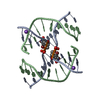
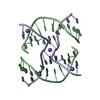
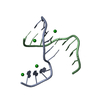
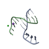
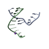
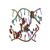
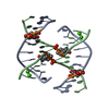
| Title | DNA Holliday junction stabilized by chlorine halogen bond. | ||||||
 Components Components |
| ||||||
 Keywords Keywords | DNA / DNA Holliday junction / halogen bond | ||||||
| Function / homology | DNA Function and homology information Function and homology information | ||||||
| Method |  X-RAY DIFFRACTION / SYNCHROTRON / MOLECULAR REPLACEMENT / Resolution: 1.94 Å X-RAY DIFFRACTION / SYNCHROTRON / MOLECULAR REPLACEMENT / Resolution: 1.94 Å | ||||||
 Authors Authors | Carter, M. / Ho, P.S. | ||||||
 Citation Citation | Journal: Biochemistry / Year: 2013 Title: Enthalpy-entropy compensation in biomolecular halogen bonds measured in DNA junctions. Authors: Carter, M. / Voth, A.R. / Scholfield, M.R. / Rummel, B. / Sowers, L.C. / Ho, P.S. | ||||||
| History |
|
- Structure visualization
| Structure viewer | Molecule: MolmilJmol/JSmol |
|---|
- Downloads & links
-Download
| PDBx/mmCIF format | 4gqd.cif.gz | 32.4 KB | Display | PDBx/mmCIF format |
|---|---|---|---|---|
| PDB format | pdb4gqd.ent.gz | 22.8 KB | Display | PDB format |
| PDBx/mmJSON format | 4gqd.json.gz | Tree view | PDBx/mmJSON format | |
| Others |  Other downloads Other downloads |
-Validation report
| Arichive directory | https://data.pdbj.org/pub/pdb/validation_reports/gq/4gqdftp://data.pdbj.org/pub/pdb/validation_reports/gq/4gqd | HTTPS FTP |
|---|
-Related structure data
| Related structure data | 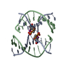 4greC 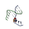 4gs2C 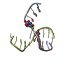 4gsgC  4gsiC 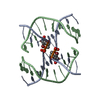 2orgS S: Starting model for refinement C: citing same article ( |
|---|---|
| Similar structure data |


-Links
 PDBj
PDBj

- Assembly
| Deposited unit | 
| |||||||||
|---|---|---|---|---|---|---|---|---|---|---|
| 1 | 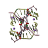
| |||||||||
| Unit cell |
| |||||||||
| Components on special symmetry positions |
| |||||||||
| Details | The biological assembly of this structure is the four stranded DNA Holliday junction. DNA Holliday junctions are generated from the two stranded asymmetric subunit through the two-fold symmetry operator at the center of the four-stranded junction. |
-Components
| #1: DNA chain | Mass: 3081.423 Da / Num. of mol.: 2 / Source method: obtained synthetically / Details: synthetically engineered DNA sequence #2: DNA chain | Mass: 3029.994 Da / Num. of mol.: 2 / Source method: obtained synthetically / Details: synthetically engineered DNA sequence #3: Chemical |   Mass: 22.990 Da / Num. of mol.: 3 / Source method: obtained synthetically / Formula: Na Mass: 22.990 Da / Num. of mol.: 3 / Source method: obtained synthetically / Formula: Na#4: Water | ChemComp-HOH / |  Mass: 18.015 Da / Num. of mol.: 93 / Source method: isolated from a natural source / Formula: H2O Mass: 18.015 Da / Num. of mol.: 93 / Source method: isolated from a natural source / Formula: H2O |
|---|
-Experimental details
-Experiment
| Experiment | Method: X-RAY DIFFRACTION / Number of used crystals: 1 |
|---|
- Sample preparation
| Crystal grow | Temperature: 298 K / Method: vapor diffusion, sitting drop / pH: 7 Details: 0.7mM DNA, 25mM sodium cacodylate pH 7.0 buffer, 10-25mM calcium chloride, and 0.8-1.2mM spermine, equilibrated against a reservoir of 30-40% aqueous MPD., VAPOR DIFFUSION, SITTING DROP, temperature 298K |
|---|
-Data collection
| Diffraction | Mean temperature: 200 K |
|---|---|
| Diffraction source | Source: SYNCHROTRON / Site: ALS  / Beamline: 4.2.2 / Wavelength: 0.9 Å / Beamline: 4.2.2 / Wavelength: 0.9 Å |
| Detector | Type: NOIR-1 / Detector: CCD / Date: Oct 21, 2009 |
| Radiation | Monochromator: ROSENBAUM-ROCK SI(111) SAGITALLY FOCUSED MONOCHROMATOR Protocol: SINGLE WAVELENGTH / Monochromatic (M) / Laue (L): M / Scattering type: x-ray |
| Radiation wavelength | Wavelength: 0.9 Å / Relative weight: 1 |
| Reflection | Resolution: 1.94→30.68 Å / Num. all: 4793 / Num. obs: 3538 / % possible obs: 73.8 % / Observed criterion σ(F): 1.4 / Observed criterion σ(I): 2 / Biso Wilson estimate: 4.6 Å2 |
| Reflection shell | Resolution: 1.94→2.06 Å / Redundancy: 1.02 % / Rmerge(I) obs: 0.228 / Mean I/σ(I) obs: 3.25 / Num. unique all: 638 / Rsym value: 0.268 / % possible all: 54.2 |
- Processing
| Software |
| ||||||||||||||||||||||||||||||||||||||||||||||||||||||||||||||||||||||||||||||||
|---|---|---|---|---|---|---|---|---|---|---|---|---|---|---|---|---|---|---|---|---|---|---|---|---|---|---|---|---|---|---|---|---|---|---|---|---|---|---|---|---|---|---|---|---|---|---|---|---|---|---|---|---|---|---|---|---|---|---|---|---|---|---|---|---|---|---|---|---|---|---|---|---|---|---|---|---|---|---|---|---|---|
| Refinement | Method to determine structure: MOLECULAR REPLACEMENT Starting model: 2ORG Resolution: 1.94→30.68 Å / Rfactor Rfree error: 0.023 / Data cutoff high absF: 28646.87 / Data cutoff low absF: 0 / Isotropic thermal model: RESTRAINED / Cross valid method: THROUGHOUT / σ(F): 1.4 / σ(I): 2 / Stereochemistry target values: CNS / Details: BULK SOLVENT MODEL USED
| ||||||||||||||||||||||||||||||||||||||||||||||||||||||||||||||||||||||||||||||||
| Solvent computation | Solvent model: FLAT MODEL / Bsol: 77.8587 Å2 / ksol: 0.3 e/Å3 | ||||||||||||||||||||||||||||||||||||||||||||||||||||||||||||||||||||||||||||||||
| Displacement parameters | Biso mean: 17.2 Å2
| ||||||||||||||||||||||||||||||||||||||||||||||||||||||||||||||||||||||||||||||||
| Refine analyze |
| ||||||||||||||||||||||||||||||||||||||||||||||||||||||||||||||||||||||||||||||||
| Refinement step | Cycle: LAST / Resolution: 1.94→30.68 Å
| ||||||||||||||||||||||||||||||||||||||||||||||||||||||||||||||||||||||||||||||||
| Refine LS restraints |
| ||||||||||||||||||||||||||||||||||||||||||||||||||||||||||||||||||||||||||||||||
| LS refinement shell | Resolution: 1.94→2.06 Å / Rfactor Rfree error: 0.127 / Total num. of bins used: 6
| ||||||||||||||||||||||||||||||||||||||||||||||||||||||||||||||||||||||||||||||||
| Xplor file |
|
