 Movie
Movie Controller

[English] 日本語
 Yorodumi
- PDB-3hs1: Structure of Holliday junction formed by d(CCGGTACCGG); Crystal g... -
+ Open data

- Basic information
| Entry | Database: PDB / ID: 3hs1 | ||||||||||||||||||
|---|---|---|---|---|---|---|---|---|---|---|---|---|---|---|---|---|---|---|---|
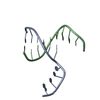
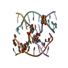
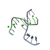
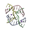
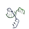
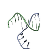
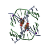
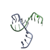
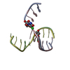
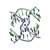
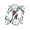
| Title | Structure of Holliday junction formed by d(CCGGTACCGG); Crystal grown with CoCl2 | ||||||||||||||||||
 Components Components | 5'-D(* Keywords KeywordsDNA / Junction with B-DNA arms | Function / homology | DNA |  Function and homology information Function and homology informationMethod |  X-RAY DIFFRACTION / MOLECULAR REPLACEMENT / Resolution: 2.51 Å X-RAY DIFFRACTION / MOLECULAR REPLACEMENT / Resolution: 2.51 Å  Authors AuthorsVenkadesh, S. / Mandal, P.K. / Gautham, N. |  CitationJournal: To be Published CitationJournal: To be PublishedTitle: Crystal structure of Holliday junction Authors: Venkadesh, S. / Mandal, P.K. / Gautham, N. History |
|
- Structure visualization
| Structure viewer | Molecule: MolmilJmol/JSmol |
|---|
- Downloads & links
-Download
| PDBx/mmCIF format | 3hs1.cif.gz | 19.9 KB | Display | PDBx/mmCIF format |
|---|---|---|---|---|
| PDB format | pdb3hs1.ent.gz | 13 KB | Display | PDB format |
| PDBx/mmJSON format | 3hs1.json.gz | Tree view | PDBx/mmJSON format | |
| Others |  Other downloads Other downloads |
-Validation report
| Arichive directory | https://data.pdbj.org/pub/pdb/validation_reports/hs/3hs1ftp://data.pdbj.org/pub/pdb/validation_reports/hs/3hs1 | HTTPS FTP |
|---|
-Related structure data
| Related structure data |  1jucS S: Starting model for refinement |
|---|---|
| Similar structure data |
-Links
 PDBj
PDBj

- Assembly
| Deposited unit | 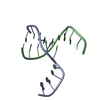
| ||||||||
|---|---|---|---|---|---|---|---|---|---|
| 1 | 
| ||||||||
| Unit cell |
| ||||||||
| Components on special symmetry positions |
|
-Components
| #1: DNA chain | Mass: 3045.992 Da / Num. of mol.: 2 / Source method: obtained synthetically #2: Water | ChemComp-HOH / |  Mass: 18.015 Da / Num. of mol.: 19 / Source method: isolated from a natural source / Formula: H2O Mass: 18.015 Da / Num. of mol.: 19 / Source method: isolated from a natural source / Formula: H2O |
|---|
-Experimental details
-Experiment
| Experiment | Method: X-RAY DIFFRACTION / Number of used crystals: 1 |
|---|
- Sample preparation
| Crystal | Density Matthews: 2.31 Å3/Da / Density % sol: 46.75 % |
|---|---|
| Crystal grow | Temperature: 293 K / Method: vapor diffusion, hanging drop / pH: 7 Details: 1mM DNA, 50mM Sodium Cacodylate buffer, 1mM CoCl2, 10mM spermine, 50% MPD, pH 7.0, VAPOR DIFFUSION, HANGING DROP, temperature 293K |
-Data collection
| Diffraction | Mean temperature: 100 K |
|---|---|
| Diffraction source | Source: ROTATING ANODE / Type: BRUKER AXS MICROSTAR / Wavelength: 1.5418 Å |
| Detector | Type: MARRESEARCH / Detector: IMAGE PLATE / Date: Feb 5, 2009 / Details: Mirrors |
| Radiation | Monochromator: Mirrors / Protocol: SINGLE WAVELENGTH / Monochromatic (M) / Laue (L): M / Scattering type: x-ray |
| Radiation wavelength | Wavelength: 1.5418 Å / Relative weight: 1 |
| Reflection | Resolution: 2.5→28.44 Å / Num. all: 1893 / Num. obs: 1864 / % possible obs: 92.9 % / Observed criterion σ(I): 1 / Redundancy: 2.84 % / Biso Wilson estimate: 63.2 Å2 / Rmerge(I) obs: 0.096 / Rsym value: 0.075 / Net I/σ(I): 4.1 |
| Reflection shell | Resolution: 2.5→2.6 Å / Redundancy: 3.08 % / Rmerge(I) obs: 0.304 / Mean I/σ(I) obs: 1.4 / Num. unique all: 172 / Rsym value: 0.262 / % possible all: 90.1 |
- Processing
| Software |
| ||||||||||||||||||||||||||||||||||||||||||||||||||||||||||||
|---|---|---|---|---|---|---|---|---|---|---|---|---|---|---|---|---|---|---|---|---|---|---|---|---|---|---|---|---|---|---|---|---|---|---|---|---|---|---|---|---|---|---|---|---|---|---|---|---|---|---|---|---|---|---|---|---|---|---|---|---|---|
| Refinement | Method to determine structure: MOLECULAR REPLACEMENT Starting model: PDB ID 1JUC Resolution: 2.51→28.44 Å / Cor.coef. Fo:Fc: 0.93 / Cor.coef. Fo:Fc free: 0.935 / SU B: 10.977 / SU ML: 0.22 / Isotropic thermal model: ISOTROPIC / Cross valid method: THROUGHOUT / ESU R: 1.671 / ESU R Free: 0.327 / Stereochemistry target values: MAXIMUM LIKELIHOOD
| ||||||||||||||||||||||||||||||||||||||||||||||||||||||||||||
| Solvent computation | Ion probe radii: 0.8 Å / Shrinkage radii: 0.8 Å / VDW probe radii: 1.2 Å / Solvent model: MASK | ||||||||||||||||||||||||||||||||||||||||||||||||||||||||||||
| Displacement parameters | Biso mean: 33.891 Å2
| ||||||||||||||||||||||||||||||||||||||||||||||||||||||||||||
| Refine analyze | Luzzati coordinate error obs: 0.472 Å | ||||||||||||||||||||||||||||||||||||||||||||||||||||||||||||
| Refinement step | Cycle: LAST / Resolution: 2.51→28.44 Å
| ||||||||||||||||||||||||||||||||||||||||||||||||||||||||||||
| Refine LS restraints |
| ||||||||||||||||||||||||||||||||||||||||||||||||||||||||||||
| LS refinement shell | Resolution: 2.506→2.57 Å / Rfactor Rfree error: 0.327 / Total num. of bins used: 20
|
