 Movie
Movie Controller

[English] 日本語
 Yorodumi
- PDB-1s1l: Influence of Groove Interactions on the Formation of DNA Holliday... -
+ Open data

- Basic information
| Entry | Database: PDB / ID: 1s1l | ||||||||||||||||||||
|---|---|---|---|---|---|---|---|---|---|---|---|---|---|---|---|---|---|---|---|---|---|
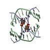
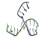
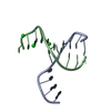
| Title | Influence of Groove Interactions on the Formation of DNA Holliday Junctions | ||||||||||||||||||||
 Components Components | 5'-D(* Keywords KeywordsDNA / Holliday Junction / DNA Four-Way Junction / major groove / Inosine / minor groove | Function / homology | DNA |  Function and homology information Function and homology informationMethod |  X-RAY DIFFRACTION / MOLECULAR REPLACEMENT / Resolution: 2.2 Å X-RAY DIFFRACTION / MOLECULAR REPLACEMENT / Resolution: 2.2 Å  Authors AuthorsHays, F.A. / Jones, Z.J. / Ho, P.S. |  CitationJournal: Biochemistry / Year: 2004 CitationJournal: Biochemistry / Year: 2004Title: Influence of minor groove substituents on the structure of DNA holliday junctions. Authors: Hays, F.A. / Jones, Z.J. / Ho, P.S. History |
Remark 300 | BIOMOLECULE: 1 THIS ENTRY CONTAINS THE CRYSTALLOGRAPHIC ASYMMETRIC UNIT WHICH CONSISTS OF 2 CHAIN(S) ...BIOMOLECULE: 1 THIS ENTRY CONTAINS THE CRYSTALLOGRAPHIC ASYMMETRIC UNIT WHICH CONSISTS OF 2 CHAIN(S). THE FULL BIOLOGICAL UNIT CONSISTS OF A FOUR STRANDED DNA HOLLIDAY JUNCTION. SEE REMARK 350 FOR INFORMATION ON GENERATING THE BIOLOGICAL MOLECULE(S). | |
- Structure visualization
| Structure viewer | Molecule: MolmilJmol/JSmol |
|---|
- Downloads & links
-Download
| PDBx/mmCIF format | 1s1l.cif.gz | 21.2 KB | Display | PDBx/mmCIF format |
|---|---|---|---|---|
| PDB format | pdb1s1l.ent.gz | 13.7 KB | Display | PDB format |
| PDBx/mmJSON format | 1s1l.json.gz | Tree view | PDBx/mmJSON format | |
| Others |  Other downloads Other downloads |
-Validation report
| Arichive directory | https://data.pdbj.org/pub/pdb/validation_reports/s1/1s1lftp://data.pdbj.org/pub/pdb/validation_reports/s1/1s1l | HTTPS FTP |
|---|
-Related structure data
| Related structure data | 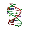 1s1kC C: citing same article ( |
|---|---|
| Similar structure data | |
| Other databases |
|


-Links
 PDBj
PDBj

- Assembly
| Deposited unit | 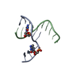
| ||||||||
|---|---|---|---|---|---|---|---|---|---|
| 1 | 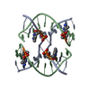
| ||||||||
| Unit cell |
| ||||||||
| Components on special symmetry positions |
| ||||||||
| Details | Full four stranded Holliday junction structure is generated by a 2-fold symmetry axis. |
-Components
| #1: DNA chain | Mass: 3045.006 Da / Num. of mol.: 2 / Source method: obtained synthetically #2: Water | ChemComp-HOH / |  Mass: 18.015 Da / Num. of mol.: 62 / Source method: isolated from a natural source / Formula: H2O Mass: 18.015 Da / Num. of mol.: 62 / Source method: isolated from a natural source / Formula: H2O |
|---|
-Experimental details
-Experiment
| Experiment | Method: X-RAY DIFFRACTION / Number of used crystals: 1 |
|---|
- Sample preparation
| Crystal | Density Matthews: 2.17 Å3/Da / Density % sol: 42.8 % | ||||||||||||||||||||||||||||
|---|---|---|---|---|---|---|---|---|---|---|---|---|---|---|---|---|---|---|---|---|---|---|---|---|---|---|---|---|---|
| Crystal grow | Method: vapor diffusion, sitting drop / pH: 7 Details: 25mM Na Cacodylate, .5mM DNA, 180mM CaCl2, 2.4mM spermine against 20% resevoir MPD, pH 7.0, VAPOR DIFFUSION, SITTING DROP | ||||||||||||||||||||||||||||
| Components of the solutions |
|
-Data collection
| Diffraction | Mean temperature: 103 K |
|---|---|
| Diffraction source | Source: ROTATING ANODE / Type: RIGAKU / Wavelength: 1.542 Å |
| Detector | Type: RIGAKU RAXIS IV / Detector: IMAGE PLATE / Date: Nov 8, 2003 / Details: osmic mirrors |
| Radiation | Protocol: SINGLE WAVELENGTH / Monochromatic (M) / Laue (L): M / Scattering type: x-ray |
| Radiation wavelength | Wavelength: 1.542 Å / Relative weight: 1 |
| Reflection | Resolution: 2→18.2 Å / Num. all: 3492 / Num. obs: 3492 / % possible obs: 93.4 % / Observed criterion σ(I): 0 / Biso Wilson estimate: 11.2 Å2 / Rmerge(I) obs: 0.05 |
| Reflection shell | Resolution: 2→2.07 Å / Rmerge(I) obs: 0.291 / Mean I/σ(I) obs: 2.4 / % possible all: 61.8 |
- Processing
| Software |
| ||||||||||||||||||||||||||||||||||||||||||||||||||||||||||||
|---|---|---|---|---|---|---|---|---|---|---|---|---|---|---|---|---|---|---|---|---|---|---|---|---|---|---|---|---|---|---|---|---|---|---|---|---|---|---|---|---|---|---|---|---|---|---|---|---|---|---|---|---|---|---|---|---|---|---|---|---|---|
| Refinement | Method to determine structure: MOLECULAR REPLACEMENT Starting model: ndb id UD0019 Resolution: 2.2→18.2 Å / Rfactor Rfree error: 0.015 / Data cutoff high absF: 44978.75 / Data cutoff high rms absF: 44978.75 / Data cutoff low absF: 0 / Isotropic thermal model: RESTRAINED / Cross valid method: THROUGHOUT / σ(F): 0 / σ(I): 0
| ||||||||||||||||||||||||||||||||||||||||||||||||||||||||||||
| Solvent computation | Solvent model: FLAT MODEL / Bsol: 50.3728 Å2 / ksol: 0.360393 e/Å3 | ||||||||||||||||||||||||||||||||||||||||||||||||||||||||||||
| Displacement parameters | Biso mean: 11.2 Å2
| ||||||||||||||||||||||||||||||||||||||||||||||||||||||||||||
| Refine analyze |
| ||||||||||||||||||||||||||||||||||||||||||||||||||||||||||||
| Refinement step | Cycle: LAST / Resolution: 2.2→18.2 Å
| ||||||||||||||||||||||||||||||||||||||||||||||||||||||||||||
| Refine LS restraints |
| ||||||||||||||||||||||||||||||||||||||||||||||||||||||||||||
| LS refinement shell | Resolution: 2.2→2.34 Å / Rfactor Rfree error: 0.053 / Total num. of bins used: 6
| ||||||||||||||||||||||||||||||||||||||||||||||||||||||||||||
| Xplor file |
|
