[English] 日本語
Yorodumi
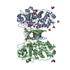
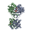
- PDB-1zcz: Crystal structure of Phosphoribosylaminoimidazolecarboxamide form... -

+
Open data


ID or keywords:

Loading...

-
Basic information

Entry
Database: PDB / ID: 1zcz
TitleCrystal structure of Phosphoribosylaminoimidazolecarboxamide formyltransferase / IMP cyclohydrolase (TM1249) from THERMOTOGA MARITIMA at 1.88 A resolution
ComponentsBifunctional purine biosynthesis protein purH
KeywordsTRANSFERASE/HYDROLASE / TM1249 / Phosphoribosylaminoimidazolecarboxamide formyltransferase (EC 2.1.2.3) / IMP cyclohydrolase (EC 3.5.4.10) / Structural Genomics / Joint Center for Structural Genomics / JCSG / Protein Structure Initiative / PSI / TRANSFERASE-HYDROLASE COMPLEX
Function / homology
Function and homology information


phosphoribosylaminoimidazolecarboxamide formyltransferase / phosphoribosylaminoimidazolecarboxamide formyltransferase activity / IMP cyclohydrolase / IMP cyclohydrolase activity / 'de novo' IMP biosynthetic process / cytosol
Similarity search - Function
AICAR transformylase, duplication domain / AICAR transformylase, duplicated domain superfamily / AICARFT/IMPCHase bienzyme / Bifunctional purine biosynthesis protein PurH-like / AICARFT/IMPCHase bienzyme / Methylglyoxal synthase-like domain / MGS-like domain profile. / Methylglyoxal synthase-like domain / Methylglyoxal synthase-like domain superfamily / MGS-like domain ...AICAR transformylase, duplication domain / AICAR transformylase, duplicated domain superfamily / AICARFT/IMPCHase bienzyme / Bifunctional purine biosynthesis protein PurH-like / AICARFT/IMPCHase bienzyme / Methylglyoxal synthase-like domain / MGS-like domain profile. / Methylglyoxal synthase-like domain / Methylglyoxal synthase-like domain superfamily / MGS-like domain / MGS-like domain / Cytidine Deaminase; domain 2 / Cytidine deaminase-like / Rossmann fold / 3-Layer(aba) Sandwich / Alpha Beta
Similarity search - Domain/homology
: / Bifunctional purine biosynthesis protein PurH
Similarity search - Component
Biological speciesThermotoga maritima (bacteria)
MethodX-RAY DIFFRACTION / SYNCHROTRON / MOLECULAR REPLACEMENT / molecular replacement / Resolution: 1.88 Å
AuthorsJoint Center for Structural Genomics (JCSG)
CitationJournal: Proteins / Year: 2008
Title: Crystal structure of AICAR transformylase IMP cyclohydrolase (TM1249) from Thermotoga maritima at 1.88 A resolution.
Authors: Axelrod, H.L. / McMullan, D. / Krishna, S.S. / Miller, M.D. / Elsliger, M.A. / Abdubek, P. / Ambing, E. / Astakhova, T. / Carlton, D. / Chiu, H.J. / Clayton, T. / Duan, L. / Feuerhelm, J. / ...Authors: Axelrod, H.L. / McMullan, D. / Krishna, S.S. / Miller, M.D. / Elsliger, M.A. / Abdubek, P. / Ambing, E. / Astakhova, T. / Carlton, D. / Chiu, H.J. / Clayton, T. / Duan, L. / Feuerhelm, J. / Grzechnik, S.K. / Hale, J. / Han, G.W. / Haugen, J. / Jaroszewski, L. / Jin, K.K. / Klock, H.E. / Knuth, M.W. / Koesema, E. / Morse, A.T. / Nigoghossian, E. / Okach, L. / Oommachen, S. / Paulsen, J. / Quijano, K. / Reyes, R. / Rife, C.L. / van den Bedem, H. / Weekes, D. / White, A. / Wolf, G. / Xu, Q. / Hodgson, K.O. / Wooley, J. / Deacon, A.M. / Godzik, A. / Lesley, S.A. / Wilson, I.A.
History
DepositionApr 13, 2005Deposition site: RCSB / Processing site: RCSB
Revision 1.0Apr 26, 2005Provider: repository / Type: Initial release
Revision 1.1Apr 30, 2008Group: Version format compliance
Revision 1.2Jul 13, 2011Group: Advisory / Version format compliance
Revision 1.3Jan 25, 2023Group: Database references / Derived calculations
Category: database_2 / pdbx_struct_conn_angle ...database_2 / pdbx_struct_conn_angle / struct_conn / struct_ref_seq_dif / struct_site
Item: _database_2.pdbx_DOI / _database_2.pdbx_database_accession ..._database_2.pdbx_DOI / _database_2.pdbx_database_accession / _pdbx_struct_conn_angle.ptnr1_auth_comp_id / _pdbx_struct_conn_angle.ptnr1_auth_seq_id / _pdbx_struct_conn_angle.ptnr1_label_atom_id / _pdbx_struct_conn_angle.ptnr1_label_comp_id / _pdbx_struct_conn_angle.ptnr1_label_seq_id / _pdbx_struct_conn_angle.ptnr3_auth_comp_id / _pdbx_struct_conn_angle.ptnr3_auth_seq_id / _pdbx_struct_conn_angle.ptnr3_label_atom_id / _pdbx_struct_conn_angle.ptnr3_label_comp_id / _pdbx_struct_conn_angle.ptnr3_label_seq_id / _pdbx_struct_conn_angle.value / _struct_conn.pdbx_dist_value / _struct_conn.ptnr1_auth_comp_id / _struct_conn.ptnr1_auth_seq_id / _struct_conn.ptnr1_label_asym_id / _struct_conn.ptnr1_label_atom_id / _struct_conn.ptnr1_label_comp_id / _struct_conn.ptnr1_label_seq_id / _struct_conn.ptnr2_auth_comp_id / _struct_conn.ptnr2_auth_seq_id / _struct_conn.ptnr2_label_asym_id / _struct_conn.ptnr2_label_atom_id / _struct_conn.ptnr2_label_comp_id / _struct_conn.ptnr2_label_seq_id / _struct_ref_seq_dif.details / _struct_site.pdbx_auth_asym_id / _struct_site.pdbx_auth_comp_id / _struct_site.pdbx_auth_seq_id
Revision 1.4Sep 20, 2023Group: Data collection / Refinement description
Category: chem_comp_atom / chem_comp_bond ...chem_comp_atom / chem_comp_bond / pdbx_initial_refinement_model / struct_ncs_dom_lim
Item: _struct_ncs_dom_lim.beg_auth_comp_id / _struct_ncs_dom_lim.end_auth_comp_id

-
Structure visualization

Structure viewerMolecule:
MolmilJmol/JSmol

Downloads & links

-
Assembly

Deposited unit
A: Bifunctional purine biosynthesis protein purH
B: Bifunctional purine biosynthesis protein purH
hetero molecules


Theoretical massNumber of molelcules
Total (without water)103,2526
Polymers102,7862
Non-polymers4674
Water11,458636
1


  • Idetical with deposited unit
  • defined by author&software
TypeNameSymmetry operationNumber
identity operation1_555x,y,z1
Buried area11720 Å2
ΔGint-45 kcal/mol
Surface area35540 Å2
MethodPISA
Unit cell
Length a, b, c (Å)62.084, 58.630, 72.749
Angle α, β, γ (deg.)99.26, 96.89, 106.13
Int Tables number1
Space group name H-MP1
Noncrystallographic symmetry (NCS)NCS domain:
IDEns-IDDetails
11A
21B
12A
22B

NCS domain segments:

Component-ID: 1 / Refine code: 4

Dom-IDEns-IDBeg auth comp-IDBeg label comp-IDEnd auth comp-IDEnd label comp-IDAuth asym-IDLabel asym-IDAuth seq-IDLabel seq-ID
11LYSLYSASPASPAA2 - 16614 - 178
21LYSLYSASPASPBB2 - 16614 - 178
12LEULEUHISHISAA167 - 452179 - 464
22LEULEUHISHISBB167 - 452179 - 464

NCS ensembles :
ID
1
2

-
Components

#1: Protein Bifunctional purine biosynthesis protein purH / Phosphoribosylaminoimidazolecarboxamide formyltransferase / IMP cyclohydrolase


Mass: 51392.867 Da / Num. of mol.: 2 / Mutation: 2.1.2.3, 3.5.4.10
Source method: isolated from a genetically manipulated source
Source: (gene. exp.) Thermotoga maritima (bacteria) / Gene: purH / Production host: Escherichia coli (E. coli) / References: UniProt: Q9X0X6
#2: Chemical ChemComp-K / POTASSIUM ION


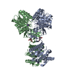
Mass: 39.098 Da / Num. of mol.: 2 / Source method: obtained synthetically / Formula: K
#3: Chemical ChemComp-PG4 / TETRAETHYLENE GLYCOL / Polyethylene glycol


Mass: 194.226 Da / Num. of mol.: 2 / Source method: obtained synthetically / Formula: C8H18O5 / Comment: precipitant*YM
#4: Water ChemComp-HOH / water / Water


Mass: 18.015 Da / Num. of mol.: 636 / Source method: isolated from a natural source / Formula: H2O

-
Experimental details

-
Experiment

ExperimentMethod: X-RAY DIFFRACTION / Number of used crystals: 1

-
Sample preparation

CrystalDensity Matthews: 2.3 Å3/Da / Density % sol: 46.12 %
Crystal growTemperature: 293 K / Method: vapor diffusion, sitting drop, nanodrop / pH: 5
Details: 20.0% PEG-6000, 0.1M Citrate pH 5.0, VAPOR DIFFUSION,SITTING DROP,NANODROP, temperature 293K

-
Data collection

DiffractionMean temperature: 100 K
Diffraction sourceSource: SYNCHROTRON / Site: ALS / Beamline: 5.0.1 / Wavelength: 1
DetectorType: ADSC QUANTUM 210 / Detector: CCD / Date: Feb 3, 2005 / Details: Single crystal, cylindrically bent, Si(220)
RadiationMonochromator: Single crystal, cylindrically bent, Si(220) / Protocol: SINGLE WAVELENGTH / Monochromatic (M) / Laue (L): M / Scattering type: x-ray
Radiation wavelengthWavelength: 1 Å / Relative weight: 1
ReflectionResolution: 1.88→50 Å / Num. obs: 74289 / % possible obs: 95.6 % / Redundancy: 2.9 % / Rsym value: 0.056 / Χ2: 0.962 / Net I/σ(I): 25.65
Reflection shell

Diffraction-ID: 1

Resolution (Å)% possible obs (%)Redundancy (%)Mean I/σ(I) obsNum. measured obsRsym valueΧ2% possible all
1.88-1.9170.61.60.020527370.3140.90570.6
1.91-1.9587.31.534080.2580.909
1.95-1.9893.61.636030.2210.925
1.98-2.0395.31.637080.1720.92
2.03-2.0796.21.737730.1450.957
2.07-2.1296.21.936750.1280.909
2.12-2.1797.1237890.130.97
2.17-2.23972.137970.1390.917
2.23-2.2997.12.237700.150.993
2.29-2.3797.42.337700.1690.953
2.37-2.4597.62.537840.1880.96
2.45-2.5597.82.838000.1680.917
2.55-2.67983.138270.1660.953
2.67-2.8198.13.538140.1380.949
2.81-2.9898.3438090.1141.003
2.98-3.2198.64.438290.081.01
3.21-3.5498.74.437990.0540.997
3.54-4.0599.14.438830.0431.006
4.05-5.199.24.438540.0350.918
5.1-5099.44.438600.0330.93

-
Phasing

PhasingMethod: molecular replacement

-
Processing

Software
NameVersionClassificationNB
REFMAC5.2.0005refinement
SCALEPACKdata scaling
PDB_EXTRACT1.6data extraction
DENZOdata reduction
RefinementMethod to determine structure: MOLECULAR REPLACEMENT
Starting model: 1m9n 1g8mA
Resolution: 1.88→50 Å / Cor.coef. Fo:Fc: 0.973 / Cor.coef. Fo:Fc free: 0.956 / SU B: 5.959 / SU ML: 0.092 / TLS residual ADP flag: LIKELY RESIDUAL / Cross valid method: THROUGHOUT / ESU R: 0.135 / ESU R Free: 0.127 / Stereochemistry target values: MAXIMUM LIKELIHOOD
Details: 1). HYDROGENS HAVE BEEN ADDED IN THE RIDING POSITIONS 2). ELECTRON DENSITY INDICATES A CIS-PEPTIDE BOND CONFORMATION BETWEEN RESIDUES 355 AND 356 ON THE A AND B CHAINS.
RfactorNum. reflection% reflectionSelection details
Rfree0.197 3700 5 %RANDOM
Rwork0.156 ---
all0.158 ---
obs0.15849 69797 95.89 %-
Solvent computationIon probe radii: 0.8 Å / Shrinkage radii: 0.8 Å / VDW probe radii: 1.2 Å / Solvent model: BABINET MODEL WITH MASK
Displacement parametersBiso mean: 36.497 Å2
Baniso -1Baniso -2Baniso -3
1--1.56 Å2-0.09 Å2-1.16 Å2
2--0.21 Å2-1.07 Å2
3---0.68 Å2
Refinement stepCycle: LAST / Resolution: 1.88→50 Å
ProteinNucleic acidLigandSolventTotal
Num. atoms6961 0 28 636 7625
Refine LS restraints
Refine-IDTypeDev idealDev ideal targetNumber
X-RAY DIFFRACTIONr_bond_refined_d0.0130.0227186
X-RAY DIFFRACTIONr_bond_other_d0.0050.026694
X-RAY DIFFRACTIONr_angle_refined_deg1.3951.9699722
X-RAY DIFFRACTIONr_angle_other_deg0.985315552
X-RAY DIFFRACTIONr_dihedral_angle_1_deg5.7855904
X-RAY DIFFRACTIONr_dihedral_angle_2_deg35.02524.257303
X-RAY DIFFRACTIONr_dihedral_angle_3_deg13.86151229
X-RAY DIFFRACTIONr_dihedral_angle_4_deg16.2511538
X-RAY DIFFRACTIONr_chiral_restr0.0870.21088
X-RAY DIFFRACTIONr_gen_planes_refined0.0060.027973
X-RAY DIFFRACTIONr_gen_planes_other0.0020.021443
X-RAY DIFFRACTIONr_nbd_refined0.220.21433
X-RAY DIFFRACTIONr_nbd_other0.1780.26592
X-RAY DIFFRACTIONr_nbtor_refined0.1770.23418
X-RAY DIFFRACTIONr_nbtor_other0.0840.24224
X-RAY DIFFRACTIONr_xyhbond_nbd_refined0.1550.2505
X-RAY DIFFRACTIONr_metal_ion_refined0.2230.28
X-RAY DIFFRACTIONr_symmetry_vdw_refined0.0710.215
X-RAY DIFFRACTIONr_symmetry_vdw_other0.2630.256
X-RAY DIFFRACTIONr_symmetry_hbond_refined0.0980.213
X-RAY DIFFRACTIONr_mcbond_it1.60334511
X-RAY DIFFRACTIONr_mcbond_other0.61531846
X-RAY DIFFRACTIONr_mcangle_it2.57757234
X-RAY DIFFRACTIONr_scbond_it4.29682698
X-RAY DIFFRACTIONr_scangle_it6.267112488
Refine LS restraints NCS

Dom-ID: 1 / Auth asym-ID: A / Refine-ID: X-RAY DIFFRACTION

Ens-IDNumberTypeRms dev position (Å)Weight position
12423medium positional0.470.5
24150medium positional0.570.5
12423medium thermal1.072
24150medium thermal0.882
LS refinement shellResolution: 1.882→1.93 Å / Total num. of bins used: 20
RfactorNum. reflection% reflection
Rfree0.297 168 -
Rwork0.217 3246 -
obs--73.74 %
Refinement TLS params.

Method: refined / Refine-ID: X-RAY DIFFRACTION

IDL112)L122)L132)L222)L232)L332)S11 (Å °)S12 (Å °)S13 (Å °)S21 (Å °)S22 (Å °)S23 (Å °)S31 (Å °)S32 (Å °)S33 (Å °)T112)T122)T132)T222)T232)T332)Origin x (Å)Origin y (Å)Origin z (Å)
12.4616-0.50320.53252.786-1.03581.9179-0.0533-0.223-0.43110.35590.10310.05640.2338-0.1339-0.0498-0.09080.01740.0298-0.0840.0176-0.153413.654-17.056222.9795
22.5794-0.46080.39911.3201-0.12420.83630.0095-0.1478-0.04790.0663-0.00070.2415-0.0087-0.1241-0.0088-0.1624-0.00940.0383-0.1630.0061-0.2-20.39571.1171-5.9379
32.494-0.00421.11234.28681.70232.9846-0.1433-0.01780.45190.29020.3506-0.8557-0.37870.3327-0.2072-0.06120.0019-0.0191-0.0625-0.045-0.045522.81145.209721.6997
41.2391-0.09110.23780.97950.19252.2714-0.06550.12840.1168-0.1472-0.0151-0.0802-0.17810.10820.0806-0.1764-0.01080.0183-0.18120.0387-0.1748-1.09455.9959-20.6128
Refinement TLS group

Refine-ID: X-RAY DIFFRACTION / Selection: ALL

IDRefine TLS-IDAuth asym-IDLabel asym-IDAuth seq-IDLabel seq-ID
11AA2 - 16614 - 178
22AA167 - 452179 - 464
33BB-1 - 16611 - 178
44BB167 - 452179 - 464

+
About Yorodumi

-
News

-
Feb 9, 2022. New format data for meta-information of EMDB entries

New format data for meta-information of EMDB entries

  • Version 3 of the EMDB header file is now the official format.
  • The previous official version 1.9 will be removed from the archive.

Related info.:EMDB header

External links:wwPDB to switch to version 3 of the EMDB data model

-
Aug 12, 2020. Covid-19 info

Covid-19 info

URL: https://pdbj.org/emnavi/covid19.php

New page: Covid-19 featured information page in EM Navigator.

Related info.:Covid-19 info / Mar 5, 2020. Novel coronavirus structure data

+
Mar 5, 2020. Novel coronavirus structure data

Novel coronavirus structure data

Related info.:Yorodumi Speices / Aug 12, 2020. Covid-19 info

External links:COVID-19 featured content - PDBj / Molecule of the Month (242):Coronavirus Proteases

+
Jan 31, 2019. EMDB accession codes are about to change! (news from PDBe EMDB page)

EMDB accession codes are about to change! (news from PDBe EMDB page)

  • The allocation of 4 digits for EMDB accession codes will soon come to an end. Whilst these codes will remain in use, new EMDB accession codes will include an additional digit and will expand incrementally as the available range of codes is exhausted. The current 4-digit format prefixed with “EMD-” (i.e. EMD-XXXX) will advance to a 5-digit format (i.e. EMD-XXXXX), and so on. It is currently estimated that the 4-digit codes will be depleted around Spring 2019, at which point the 5-digit format will come into force.
  • The EM Navigator/Yorodumi systems omit the EMD- prefix.

Related info.:Q: What is EMD? / ID/Accession-code notation in Yorodumi/EM Navigator

External links:EMDB Accession Codes are Changing Soon! / Contact to PDBj

+
Jul 12, 2017. Major update of PDB

Major update of PDB

  • wwPDB released updated PDB data conforming to the new PDBx/mmCIF dictionary.
  • This is a major update changing the version number from 4 to 5, and with Remediation, in which all the entries are updated.
  • In this update, many items about electron microscopy experimental information are reorganized (e.g. em_software).
  • Now, EM Navigator and Yorodumi are based on the updated data.

External links:wwPDB Remediation / Enriched Model Files Conforming to OneDep Data Standards Now Available in the PDB FTP Archive

-
Yorodumi

Thousand views of thousand structures

  • Yorodumi is a browser for structure data from EMDB, PDB, SASBDB, etc.
  • This page is also the successor to EM Navigator detail page, and also detail information page/front-end page for Omokage search.
  • The word "yorodu" (or yorozu) is an old Japanese word meaning "ten thousand". "mi" (miru) is to see.

Related info.:EMDB / PDB / SASBDB / Comparison of 3 databanks / Yorodumi Search / Aug 31, 2016. New EM Navigator & Yorodumi / Yorodumi Papers / Jmol/JSmol / Function and homology information / Changes in new EM Navigator and Yorodumi

Read more