 Movie
Movie Controller


+ Open data

- Basic information
| Entry | Database: PDB / ID: 1y47 | ||||||
|---|---|---|---|---|---|---|---|
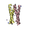
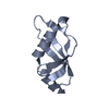
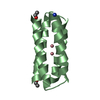
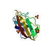
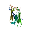
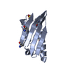
| Title | Structural studies of designed alpha-helical hairpins | ||||||
 Components Components | dueferri (DF2) | ||||||
 Keywords Keywords | DE NOVO PROTEIN / Protein design / helical hairpin / turns / diiron proteins | ||||||
| Function / homology | Immunoglobulin FC, subunit C / Single alpha-helices involved in coiled-coils or other helix-helix interfaces / Up-down Bundle / Mainly Alpha / :  Function and homology information Function and homology information | ||||||
| Method |  X-RAY DIFFRACTION / MOLECULAR REPLACEMENT / Resolution: 2.7 Å X-RAY DIFFRACTION / MOLECULAR REPLACEMENT / Resolution: 2.7 Å | ||||||
 Authors Authors | Lahr, S.J. / Engel, D.E. / Stayrook, S.E. / Maglio, O. / North, B. / Geremia, S. / Lombardi, A. / DeGrado, W.F. | ||||||
 Citation Citation | Journal: J.Mol.Biol. / Year: 2005 Title: Analysis and design of turns in alpha-helical hairpins Authors: Lahr, S.J. / Engel, D.E. / Stayrook, S.E. / Maglio, O. / North, B. / Geremia, S. / Lombardi, A. / DeGrado, W.F. | ||||||
| History |
|
- Structure visualization
| Structure viewer | Molecule: MolmilJmol/JSmol |
|---|
- Downloads & links
-Download
| PDBx/mmCIF format | 1y47.cif.gz | 31.4 KB | Display | PDBx/mmCIF format |
|---|---|---|---|---|
| PDB format | pdb1y47.ent.gz | 21.3 KB | Display | PDB format |
| PDBx/mmJSON format | 1y47.json.gz | Tree view | PDBx/mmJSON format | |
| Others |  Other downloads Other downloads |
-Validation report
| Arichive directory | https://data.pdbj.org/pub/pdb/validation_reports/y4/1y47ftp://data.pdbj.org/pub/pdb/validation_reports/y4/1y47 | HTTPS FTP |
|---|
-Related structure data
| Related structure data | 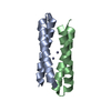 1mftC 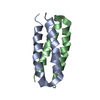 1u7jC  1u7mC 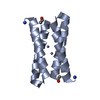 1ec5S S: Starting model for refinement C: citing same article ( |
|---|---|
| Similar structure data |


-Links
 PDBj
PDBj
- Assembly
| Deposited unit | 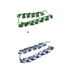
| |||||||||
|---|---|---|---|---|---|---|---|---|---|---|
| 1 | 
| |||||||||
| 2 | 
| |||||||||
| 3 | 
| |||||||||
| Unit cell |
| |||||||||
| Components on special symmetry positions |
| |||||||||
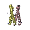
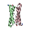
| Details | the biological assembly is a dimer generated by the two fold axis |
-Components
| #1: Protein/peptide | Mass: 5600.354 Da / Num. of mol.: 2 / Source method: obtained synthetically / Details: designed peptide #2: Chemical |   Mass: 112.411 Da / Num. of mol.: 3 / Source method: obtained synthetically / Formula: Cd Mass: 112.411 Da / Num. of mol.: 3 / Source method: obtained synthetically / Formula: Cd#3: Water | ChemComp-HOH / |  Mass: 18.015 Da / Num. of mol.: 3 / Source method: isolated from a natural source / Formula: H2O Mass: 18.015 Da / Num. of mol.: 3 / Source method: isolated from a natural source / Formula: H2O |
|---|
-Experimental details
-Experiment
| Experiment | Method: X-RAY DIFFRACTION / Number of used crystals: 1 |
|---|
- Sample preparation
| Crystal | Density Matthews: 1.7 Å3/Da / Density % sol: 31 % |
|---|---|
| Crystal grow | Temperature: 291 K / Method: vapor diffusion, hanging drop / pH: 4.6 Details: 22% PEG MW 400, 0.1 M CdCl2, 0.1 M sodium acetate pH 4.6, VAPOR DIFFUSION, HANGING DROP, temperature 291K |
-Data collection
| Diffraction | Mean temperature: 100 K |
|---|---|
| Diffraction source | Source: ROTATING ANODE / Type: RIGAKU RU300 / Wavelength: 1.5418 Å |
| Detector | Type: MARRESEARCH / Detector: IMAGE PLATE / Date: Apr 23, 2001 / Details: Osmic mirrors |
| Radiation | Monochromator: Osmic Mirrors / Protocol: SINGLE WAVELENGTH / Monochromatic (M) / Laue (L): M / Scattering type: x-ray |
| Radiation wavelength | Wavelength: 1.5418 Å / Relative weight: 1 |
| Reflection | Resolution: 2.7→15 Å / Num. all: 2640 / Num. obs: 2625 / % possible obs: 99.7 % / Observed criterion σ(F): 0 / Observed criterion σ(I): 0 / Redundancy: 10.5 % / Biso Wilson estimate: 86.1 Å2 / Rmerge(I) obs: 0.08 / Rsym value: 0.08 |
| Reflection shell | Resolution: 2.7→2.87 Å / Redundancy: 9.6 % / Rmerge(I) obs: 0.365 / Num. unique all: 379 / % possible all: 97.1 |
- Processing
| Software |
| ||||||||||||||||||||||||||||||||||||
|---|---|---|---|---|---|---|---|---|---|---|---|---|---|---|---|---|---|---|---|---|---|---|---|---|---|---|---|---|---|---|---|---|---|---|---|---|---|
| Refinement | Method to determine structure: MOLECULAR REPLACEMENT Starting model: PDB ENTRY 1EC5 Resolution: 2.7→15 Å / Rfactor Rfree error: 0.02 / Data cutoff high absF: 492107.92 / Data cutoff low absF: 0 / Isotropic thermal model: RESTRAINED / Cross valid method: THROUGHOUT / σ(F): 0 / Stereochemistry target values: Engh & Huber
| ||||||||||||||||||||||||||||||||||||
| Solvent computation | Solvent model: FLAT MODEL / Bsol: 83.7173 Å2 / ksol: 0.468996 e/Å3 | ||||||||||||||||||||||||||||||||||||
| Displacement parameters | Biso mean: 62 Å2
| ||||||||||||||||||||||||||||||||||||
| Refine analyze |
| ||||||||||||||||||||||||||||||||||||
| Refinement step | Cycle: LAST / Resolution: 2.7→15 Å
| ||||||||||||||||||||||||||||||||||||
| Refine LS restraints |
| ||||||||||||||||||||||||||||||||||||
| LS refinement shell | Resolution: 2.7→2.87 Å / Rfactor Rfree error: 0.062 / Total num. of bins used: 6
| ||||||||||||||||||||||||||||||||||||
| Xplor file |
|
