 ムービー
ムービー コントローラー


+ データを開く

- 基本情報
基本情報
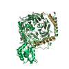
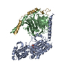
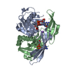
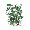
| 登録情報 | データベース: PDB / ID: 1xhm | ||||||
|---|---|---|---|---|---|---|---|
| タイトル | The Crystal Structure of a Biologically Active Peptide (SIGK) Bound to a G Protein Beta:Gamma Heterodimer | ||||||
 要素 要素 |
| ||||||
 キーワード キーワード | SIGNALING PROTEIN / WD40 Repeat / Beta-Propeller / Protein-Peptide Complex | ||||||
| 機能・相同性 |  機能・相同性情報 機能・相同性情報Olfactory Signaling Pathway / Sensory perception of sweet, bitter, and umami (glutamate) taste / Synthesis, secretion, and inactivation of Glucagon-like Peptide-1 (GLP-1) / Activation of the phototransduction cascade / Activation of G protein gated Potassium channels / G-protein activation / G beta:gamma signalling through PI3Kgamma / Prostacyclin signalling through prostacyclin receptor / G beta:gamma signalling through PLC beta / ADP signalling through P2Y purinoceptor 1 ...Olfactory Signaling Pathway / Sensory perception of sweet, bitter, and umami (glutamate) taste / Synthesis, secretion, and inactivation of Glucagon-like Peptide-1 (GLP-1) / Activation of the phototransduction cascade / Activation of G protein gated Potassium channels / G-protein activation / G beta:gamma signalling through PI3Kgamma / Prostacyclin signalling through prostacyclin receptor / G beta:gamma signalling through PLC beta / ADP signalling through P2Y purinoceptor 1 / Thromboxane signalling through TP receptor / Presynaptic function of Kainate receptors / G beta:gamma signalling through CDC42 / Inhibition of voltage gated Ca2+ channels via Gbeta/gamma subunits / G alpha (12/13) signalling events / Glucagon-type ligand receptors / G beta:gamma signalling through BTK / ADP signalling through P2Y purinoceptor 12 / Adrenaline,noradrenaline inhibits insulin secretion / Cooperation of PDCL (PhLP1) and TRiC/CCT in G-protein beta folding / Ca2+ pathway / Thrombin signalling through proteinase activated receptors (PARs) / G alpha (z) signalling events / Extra-nuclear estrogen signaling / G alpha (s) signalling events / G alpha (q) signalling events / G alpha (i) signalling events / Glucagon-like Peptide-1 (GLP1) regulates insulin secretion / High laminar flow shear stress activates signaling by PIEZO1 and PECAM1:CDH5:KDR in endothelial cells / Vasopressin regulates renal water homeostasis via Aquaporins / photoreceptor disc membrane / cellular response to catecholamine stimulus / adenylate cyclase-activating dopamine receptor signaling pathway / cellular response to prostaglandin E stimulus / G-protein beta-subunit binding / heterotrimeric G-protein complex / sensory perception of taste / signaling receptor complex adaptor activity / retina development in camera-type eye / GTPase binding / phospholipase C-activating G protein-coupled receptor signaling pathway / cell population proliferation / G protein-coupled receptor signaling pathway / GTPase activity / synapse / protein-containing complex binding / membrane / plasma membrane / cytoplasm 類似検索 - 分子機能 | ||||||
| 生物種 |  | ||||||
| 手法 |  X線回折 / シンクロトロン / 分子置換 / 解像度: 2.7 Å X線回折 / シンクロトロン / 分子置換 / 解像度: 2.7 Å | ||||||
 データ登録者 データ登録者 | Davis, T.L. / Bonacci, T.M. / Smrcka, A.V. / Sprang, S.R. | ||||||
 引用 引用 | ジャーナル: Biochemistry / 年: 2005 タイトル: Structural and Molecular Characterization of a Preferred Protein Interaction Surface on G Protein betagamma Subunits. 著者: Davis, T.L. / Bonacci, T.M. / Sprang, S.R. / Smrcka, A.V. #1: ジャーナル: To be Publishedタイトル: Molecular Determinants on G Protein Beta:Gamma Subunits Responsible for Multiple Target Recognition and Nucleotide Exchange Independent Subunit Dissociation 著者: Bonacci, T.M. / Davis, T.L. / Sprang, S.R. / Smrcka, A.V. | ||||||
| 履歴 |
|
- 構造の表示
構造の表示
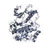
| 構造ビューア | 分子: MolmilJmol/JSmol |
|---|
- ダウンロードとリンク
-ダウンロード
| PDBx/mmCIF形式 | 1xhm.cif.gz | 90 KB | 表示 | PDBx/mmCIF形式 |
|---|---|---|---|---|
| PDB形式 | pdb1xhm.ent.gz | 67.3 KB | 表示 | PDB形式 |
| PDBx/mmJSON形式 | 1xhm.json.gz | ツリー表示 | PDBx/mmJSON形式 | |
| その他 |  その他のダウンロード その他のダウンロード |
-検証レポート
| アーカイブディレクトリ | https://data.pdbj.org/pub/pdb/validation_reports/xh/1xhmftp://data.pdbj.org/pub/pdb/validation_reports/xh/1xhm | HTTPS FTP |
|---|
-関連構造データ
| 関連構造データ | 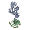 1omwS S: 精密化の開始モデル |
|---|---|
| 類似構造データ |


-リンク
 PDBj
PDBj


- 集合体
集合体
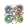
| 登録構造単位 | 
| ||||||||
|---|---|---|---|---|---|---|---|---|---|
| 1 |
| ||||||||
| 単位格子 |
|
-要素
| #1: タンパク質 | 分子量: 37416.930 Da / 分子数: 1 / 由来タイプ: 組換発現 / 由来: (組換発現) 発現宿主:   Spodoptera frugiperda (ツマジロクサヨトウ) Spodoptera frugiperda (ツマジロクサヨトウ)株 (発現宿主): High 5 / 参照: UniProt: P62871 |
|---|---|
| #2: タンパク質 | 分子量: 8690.024 Da / 分子数: 1 / 由来タイプ: 組換発現 / 由来: (組換発現) 発現宿主: Spodoptera frugiperda (ツマジロクサヨトウ)株 (発現宿主): High5 / 参照: UniProt: P63212 |
| #3: タンパク質・ペプチド | 分子量: 1688.919 Da / 分子数: 1 / 由来タイプ: 合成 詳細: The peptide was chemically synthesized. This sequence is derived from a randomized phage library. |
| #4: 水 | ChemComp-HOH /  分子量: 18.015 Da / 分子数: 37 / 由来タイプ: 天然 / 式: H2O 分子量: 18.015 Da / 分子数: 37 / 由来タイプ: 天然 / 式: H2O |
-実験情報
-実験
| 実験 | 手法: X線回折 / 使用した結晶の数: 1 |
|---|
- 試料調製
試料調製
| 結晶 | マシュー密度: 2.14 Å3/Da / 溶媒含有率: 42.1 % |
|---|---|
| 結晶化 | 温度: 293 K / 手法: 蒸気拡散法, ハンギングドロップ法 / pH: 7.5 詳細: PEG 4000, GLYCEROL, HEPES, PH 7.5, VAPOR DIFFUSION, HANGING DROP, temperature 293K |
-データ収集
| 回折 | 平均測定温度: 100 K |
|---|---|
| 放射光源 | 由来: シンクロトロン / サイト: ALS  / ビームライン: 8.2.2 / 波長: 1.0871 Å / ビームライン: 8.2.2 / 波長: 1.0871 Å |
| 検出器 | タイプ: ADSC QUANTUM 315 / 検出器: CCD / 日付: 2003年11月18日 / 詳細: mirrors |
| 放射 | モノクロメーター: DOUBLE CRYSTAL SI (111) / プロトコル: SINGLE WAVELENGTH / 単色(M)・ラウエ(L): M / 散乱光タイプ: x-ray |
| 放射波長 | 波長: 1.0871 Å / 相対比: 1 |
| 反射 | 解像度: 2.7→50 Å / Num. all: 9729 / Num. obs: 9729 / % possible obs: 88.3 % / Observed criterion σ(F): 0 / Observed criterion σ(I): -2.5 / 冗長度: 3.5 % / Biso Wilson estimate: 61.8 Å2 / Rsym value: 0.087 / Net I/σ(I): 13.5 |
| 反射 シェル | 解像度: 2.7→2.8 Å / 冗長度: 1.8 % / Rmerge(I) obs: 0.414 / Mean I/σ(I) obs: 1.6 / Num. unique all: 408 / % possible all: 38.6 |
- 解析
解析
| ソフトウェア |
| ||||||||||||||||||||||||||||||||||||||||||||||||||||||||||||
|---|---|---|---|---|---|---|---|---|---|---|---|---|---|---|---|---|---|---|---|---|---|---|---|---|---|---|---|---|---|---|---|---|---|---|---|---|---|---|---|---|---|---|---|---|---|---|---|---|---|---|---|---|---|---|---|---|---|---|---|---|---|
| 精密化 | 構造決定の手法: 分子置換 開始モデル: PDB ENTRY 1OMW 解像度: 2.7→41.91 Å / Rfactor Rfree error: 0.01 / Data cutoff high absF: 2166288.6 / Data cutoff low absF: 0 / Isotropic thermal model: GROUP / 交差検証法: THROUGHOUT / σ(F): 0 / 立体化学のターゲット値: Engh & Huber
| ||||||||||||||||||||||||||||||||||||||||||||||||||||||||||||
| 溶媒の処理 | 溶媒モデル: FLAT MODEL / Bsol: 46.0853 Å2 / ksol: 0.346454 e/Å3 | ||||||||||||||||||||||||||||||||||||||||||||||||||||||||||||
| 原子変位パラメータ | Biso mean: 46.3 Å2
| ||||||||||||||||||||||||||||||||||||||||||||||||||||||||||||
| Refine analyze |
| ||||||||||||||||||||||||||||||||||||||||||||||||||||||||||||
| 精密化ステップ | サイクル: LAST / 解像度: 2.7→41.91 Å
| ||||||||||||||||||||||||||||||||||||||||||||||||||||||||||||
| 拘束条件 |
| ||||||||||||||||||||||||||||||||||||||||||||||||||||||||||||
| LS精密化 シェル | 解像度: 2.7→2.87 Å / Rfactor Rfree error: 0.052 / Total num. of bins used: 6
| ||||||||||||||||||||||||||||||||||||||||||||||||||||||||||||
| Xplor file |
|
