 Movie
Movie Controller


+ Open data

- Basic information
| Entry | Database: PDB / ID: 1tbg | ||||||
|---|---|---|---|---|---|---|---|
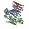
| Title | BETA-GAMMA DIMER OF THE HETEROTRIMERIC G-PROTEIN TRANSDUCIN | ||||||
 Components Components | (TRANSDUCIN) x 2 | ||||||
 Keywords Keywords | COMPLEX (GTP-BINDING/TRANSDUCER) / COMPLEX (GTP-BINDING-TRANSDUCER) / EYE / TRANSDUCER / PRENYLATION / COMPLEX (GTP-BINDING-TRANSDUCER) complex | ||||||
| Function / homology |  Function and homology information Function and homology informationOlfactory Signaling Pathway / Sensory perception of sweet, bitter, and umami (glutamate) taste / Synthesis, secretion, and inactivation of Glucagon-like Peptide-1 (GLP-1) / eye photoreceptor cell development / Inactivation, recovery and regulation of the phototransduction cascade / Activation of the phototransduction cascade / Activation of G protein gated Potassium channels / G-protein activation / G beta:gamma signalling through PI3Kgamma / Prostacyclin signalling through prostacyclin receptor ...Olfactory Signaling Pathway / Sensory perception of sweet, bitter, and umami (glutamate) taste / Synthesis, secretion, and inactivation of Glucagon-like Peptide-1 (GLP-1) / eye photoreceptor cell development / Inactivation, recovery and regulation of the phototransduction cascade / Activation of the phototransduction cascade / Activation of G protein gated Potassium channels / G-protein activation / G beta:gamma signalling through PI3Kgamma / Prostacyclin signalling through prostacyclin receptor / G beta:gamma signalling through PLC beta / ADP signalling through P2Y purinoceptor 1 / Thromboxane signalling through TP receptor / Presynaptic function of Kainate receptors / G beta:gamma signalling through CDC42 / Inhibition of voltage gated Ca2+ channels via Gbeta/gamma subunits / G alpha (12/13) signalling events / Glucagon-type ligand receptors / G beta:gamma signalling through BTK / ADP signalling through P2Y purinoceptor 12 / Adrenaline,noradrenaline inhibits insulin secretion / Cooperation of PDCL (PhLP1) and TRiC/CCT in G-protein beta folding / Ca2+ pathway / G alpha (z) signalling events / Thrombin signalling through proteinase activated receptors (PARs) / Extra-nuclear estrogen signaling / G alpha (s) signalling events / G alpha (q) signalling events / Glucagon-like Peptide-1 (GLP1) regulates insulin secretion / G alpha (i) signalling events / High laminar flow shear stress activates signaling by PIEZO1 and PECAM1:CDH5:KDR in endothelial cells / Vasopressin regulates renal water homeostasis via Aquaporins / phototransduction / intracellular protein localization / photoreceptor disc membrane / cellular response to catecholamine stimulus / adenylate cyclase-activating dopamine receptor signaling pathway / cellular response to prostaglandin E stimulus / heterotrimeric G-protein complex / G-protein beta-subunit binding / sensory perception of taste / signaling receptor complex adaptor activity / retina development in camera-type eye / GTPase binding / phospholipase C-activating G protein-coupled receptor signaling pathway / cell population proliferation / G protein-coupled receptor signaling pathway / GTPase activity / synapse / protein-containing complex binding / membrane / cytoplasm Similarity search - Function | ||||||
| Biological species |  | ||||||
| Method |  X-RAY DIFFRACTION / SYNCHROTRON / MAD / Resolution: 2.1 Å X-RAY DIFFRACTION / SYNCHROTRON / MAD / Resolution: 2.1 Å | ||||||
 Authors Authors | Sondek, J.S. / Bohm, A. / Lambright, D.G. / Hamm, H.E. / Sigler, P.B. | ||||||
 Citation Citation | Journal: Nature / Year: 1996 Title: Crystal structure of a G-protein beta gamma dimer at 2.1A resolution. Authors: Sondek, J. / Bohm, A. / Lambright, D.G. / Hamm, H.E. / Sigler, P.B. #1: Journal: Nature / Year: 1996Title: The 2.0 A Crystal Structure of a Heterotrimeric G Protein Authors: Lambright, D.G. / Sondek, J. / Bohm, A. / Skiba, N.P. / Hamm, H.E. / Sigler, P.B. #2: Journal: Nature / Year: 1994Title: Structural Determinants for Activation of the Alpha-Subunit of a Heterotrimeric G Protein Authors: Lambright, D.G. / Noel, J.P. / Hamm, H.E. / Sigler, P.B. #3: Journal: Nature / Year: 1994Title: Gtpase Mechanism of Gproteins from the 1.7-A Crystal Structure of Transducin Alpha-Gdp-Aif-4 Authors: Sondek, J. / Lambright, D.G. / Noel, J.P. / Hamm, H.E. / Sigler, P.B. #4: Journal: Nature / Year: 1993Title: The 2.2 A Crystal Structure of Transducin-Alpha Complexed with GTP Gamma S Authors: Noel, J.P. / Hamm, H.E. / Sigler, P.B. | ||||||
| History |
|
- Structure visualization
| Structure viewer | Molecule: MolmilJmol/JSmol |
|---|
- Downloads & links
-Download
| PDBx/mmCIF format | 1tbg.cif.gz | 326.9 KB | Display | PDBx/mmCIF format |
|---|---|---|---|---|
| PDB format | pdb1tbg.ent.gz | 269.7 KB | Display | PDB format |
| PDBx/mmJSON format | 1tbg.json.gz | Tree view | PDBx/mmJSON format | |
| Others |  Other downloads Other downloads |
-Validation report
| Arichive directory | https://data.pdbj.org/pub/pdb/validation_reports/tb/1tbgftp://data.pdbj.org/pub/pdb/validation_reports/tb/1tbg | HTTPS FTP |
|---|
-Related structure data
| Similar structure data |
|---|


-Links
 PDBj
PDBj


- Assembly
| Deposited unit | 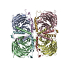
| ||||||||||||||||
|---|---|---|---|---|---|---|---|---|---|---|---|---|---|---|---|---|---|
| 1 |
| ||||||||||||||||
| 2 | 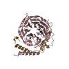
| ||||||||||||||||
| 3 | 
| ||||||||||||||||
| 4 | 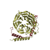
| ||||||||||||||||
| 5 | 
| ||||||||||||||||
| Unit cell |
| ||||||||||||||||
| Noncrystallographic symmetry (NCS) | NCS oper:
|
-Components
| #1: Protein | Mass: 37416.930 Da / Num. of mol.: 4 / Fragment: BETA-1 SUBUNIT / Source method: isolated from a natural source / Source: (natural) #2: Protein | Mass: 7980.186 Da / Num. of mol.: 4 / Fragment: GAMMA-1 SUBUNIT / Source method: isolated from a natural source / Source: (natural) #3: Water | ChemComp-HOH / |  Mass: 18.015 Da / Num. of mol.: 732 / Source method: isolated from a natural source / Formula: H2O Mass: 18.015 Da / Num. of mol.: 732 / Source method: isolated from a natural source / Formula: H2O |
|---|
-Experimental details
-Experiment
| Experiment | Method: X-RAY DIFFRACTION |
|---|
- Sample preparation
| Crystal | Density Matthews: 2.14 Å3/Da / Density % sol: 42.63 % | ||||||||||||||||||||||||||||||||||||||||||||||||||||||||||||||||||||||||||||||
|---|---|---|---|---|---|---|---|---|---|---|---|---|---|---|---|---|---|---|---|---|---|---|---|---|---|---|---|---|---|---|---|---|---|---|---|---|---|---|---|---|---|---|---|---|---|---|---|---|---|---|---|---|---|---|---|---|---|---|---|---|---|---|---|---|---|---|---|---|---|---|---|---|---|---|---|---|---|---|---|
| Crystal grow | Temperature: 277 K / Method: vapor diffusion, hanging drop / pH: 4.2 Details: 15-20 MG/ML PROTEIN MIXED 1:1 WITH WELL SOLUTION (10MM FUMARATE, PH 4.2, 10MM MGSO4, 2MM GDCL3, 5% W/V GLYCEROL, 5% W/V PEG 4000, 15MM BETA-MERCAPTOETHANOL. MIXTURE EQUILIBRATED VS. WELL ...Details: 15-20 MG/ML PROTEIN MIXED 1:1 WITH WELL SOLUTION (10MM FUMARATE, PH 4.2, 10MM MGSO4, 2MM GDCL3, 5% W/V GLYCEROL, 5% W/V PEG 4000, 15MM BETA-MERCAPTOETHANOL. MIXTURE EQUILIBRATED VS. WELL SOLUTION IN HANGING DROPS AT 4 DEGREES CELSIUS., vapor diffusion - hanging drop, temperature 277K | ||||||||||||||||||||||||||||||||||||||||||||||||||||||||||||||||||||||||||||||
| Crystal grow | *PLUS Temperature: 4 ℃ / pH: 7.5 / Method: vapor diffusion, hanging dropDetails: protein solution is mixed in a 1:1 ratio with well solution | ||||||||||||||||||||||||||||||||||||||||||||||||||||||||||||||||||||||||||||||
| Components of the solutions | *PLUS
|
-Data collection
| Diffraction source | Source: SYNCHROTRON / Site: NSLS  / Beamline: X25 / Wavelength: 0.95 / Beamline: X25 / Wavelength: 0.95 |
|---|---|
| Detector | Type: FUJI / Detector: IMAGE PLATE / Date: Jun 7, 1994 |
| Radiation | Monochromatic (M) / Laue (L): M / Scattering type: x-ray |
| Radiation wavelength | Wavelength: 0.95 Å / Relative weight: 1 |
| Reflection | Num. obs: 88015 / % possible obs: 96 % / Redundancy: 2.8 % / Rmerge(I) obs: 0.088 |
| Reflection | *PLUS Highest resolution: 2.1 Å / Num. measured all: 258531 |
- Processing
| Software |
| ||||||||||||||||||||||||||||||||||||||||||||||||||||||||||||
|---|---|---|---|---|---|---|---|---|---|---|---|---|---|---|---|---|---|---|---|---|---|---|---|---|---|---|---|---|---|---|---|---|---|---|---|---|---|---|---|---|---|---|---|---|---|---|---|---|---|---|---|---|---|---|---|---|---|---|---|---|---|
| Refinement | Method to determine structure: MAD / Resolution: 2.1→6 Å / σ(F): 2
| ||||||||||||||||||||||||||||||||||||||||||||||||||||||||||||
| Displacement parameters | Biso mean: 36.4 Å2 | ||||||||||||||||||||||||||||||||||||||||||||||||||||||||||||
| Refinement step | Cycle: LAST / Resolution: 2.1→6 Å
| ||||||||||||||||||||||||||||||||||||||||||||||||||||||||||||
| Refine LS restraints |
| ||||||||||||||||||||||||||||||||||||||||||||||||||||||||||||
| Software | *PLUS Name: X-PLOR / Classification: refinement | ||||||||||||||||||||||||||||||||||||||||||||||||||||||||||||
| Refinement | *PLUS Rfactor obs: 0.2 | ||||||||||||||||||||||||||||||||||||||||||||||||||||||||||||
| Solvent computation | *PLUS | ||||||||||||||||||||||||||||||||||||||||||||||||||||||||||||
| Displacement parameters | *PLUS | ||||||||||||||||||||||||||||||||||||||||||||||||||||||||||||
| Refine LS restraints | *PLUS
|
