 Movie
Movie Controller

[English] 日本語
 Yorodumi
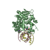
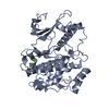
- PDB-1xhm: The Crystal Structure of a Biologically Active Peptide (SIGK) Bou... -
+ Open data

- Basic information
| Entry | Database: PDB / ID: 1xhm | ||||||
|---|---|---|---|---|---|---|---|
| Title | The Crystal Structure of a Biologically Active Peptide (SIGK) Bound to a G Protein Beta:Gamma Heterodimer | ||||||
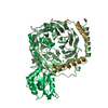
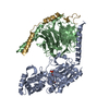
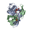
 Components Components |
| ||||||
 Keywords Keywords | SIGNALING PROTEIN / WD40 Repeat / Beta-Propeller / Protein-Peptide Complex | ||||||
| Function / homology |  Function and homology information Function and homology informationOlfactory Signaling Pathway / Sensory perception of sweet, bitter, and umami (glutamate) taste / Synthesis, secretion, and inactivation of Glucagon-like Peptide-1 (GLP-1) / Activation of the phototransduction cascade / Activation of G protein gated Potassium channels / G-protein activation / G beta:gamma signalling through PI3Kgamma / Prostacyclin signalling through prostacyclin receptor / G beta:gamma signalling through PLC beta / ADP signalling through P2Y purinoceptor 1 ...Olfactory Signaling Pathway / Sensory perception of sweet, bitter, and umami (glutamate) taste / Synthesis, secretion, and inactivation of Glucagon-like Peptide-1 (GLP-1) / Activation of the phototransduction cascade / Activation of G protein gated Potassium channels / G-protein activation / G beta:gamma signalling through PI3Kgamma / Prostacyclin signalling through prostacyclin receptor / G beta:gamma signalling through PLC beta / ADP signalling through P2Y purinoceptor 1 / Thromboxane signalling through TP receptor / Presynaptic function of Kainate receptors / G beta:gamma signalling through CDC42 / Inhibition of voltage gated Ca2+ channels via Gbeta/gamma subunits / G alpha (12/13) signalling events / Glucagon-type ligand receptors / G beta:gamma signalling through BTK / ADP signalling through P2Y purinoceptor 12 / Adrenaline,noradrenaline inhibits insulin secretion / Cooperation of PDCL (PhLP1) and TRiC/CCT in G-protein beta folding / Ca2+ pathway / G alpha (z) signalling events / Thrombin signalling through proteinase activated receptors (PARs) / Extra-nuclear estrogen signaling / G alpha (s) signalling events / G alpha (q) signalling events / Glucagon-like Peptide-1 (GLP1) regulates insulin secretion / G alpha (i) signalling events / High laminar flow shear stress activates signaling by PIEZO1 and PECAM1:CDH5:KDR in endothelial cells / Vasopressin regulates renal water homeostasis via Aquaporins / photoreceptor disc membrane / cellular response to catecholamine stimulus / adenylate cyclase-activating dopamine receptor signaling pathway / cellular response to prostaglandin E stimulus / heterotrimeric G-protein complex / G-protein beta-subunit binding / sensory perception of taste / signaling receptor complex adaptor activity / retina development in camera-type eye / GTPase binding / phospholipase C-activating G protein-coupled receptor signaling pathway / cell population proliferation / G protein-coupled receptor signaling pathway / GTPase activity / synapse / protein-containing complex binding / membrane / plasma membrane / cytoplasm Similarity search - Function | ||||||
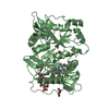
| Biological species |  | ||||||
| Method |  X-RAY DIFFRACTION / SYNCHROTRON / MOLECULAR REPLACEMENT / Resolution: 2.7 Å X-RAY DIFFRACTION / SYNCHROTRON / MOLECULAR REPLACEMENT / Resolution: 2.7 Å | ||||||
 Authors Authors | Davis, T.L. / Bonacci, T.M. / Smrcka, A.V. / Sprang, S.R. | ||||||
 Citation Citation | Journal: Biochemistry / Year: 2005 Title: Structural and Molecular Characterization of a Preferred Protein Interaction Surface on G Protein betagamma Subunits. Authors: Davis, T.L. / Bonacci, T.M. / Sprang, S.R. / Smrcka, A.V. #1: Journal: To be PublishedTitle: Molecular Determinants on G Protein Beta:Gamma Subunits Responsible for Multiple Target Recognition and Nucleotide Exchange Independent Subunit Dissociation Authors: Bonacci, T.M. / Davis, T.L. / Sprang, S.R. / Smrcka, A.V. | ||||||
| History |
|
- Structure visualization
| Structure viewer | Molecule: MolmilJmol/JSmol |
|---|
- Downloads & links
-Download
| PDBx/mmCIF format | 1xhm.cif.gz | 90 KB | Display | PDBx/mmCIF format |
|---|---|---|---|---|
| PDB format | pdb1xhm.ent.gz | 67.3 KB | Display | PDB format |
| PDBx/mmJSON format | 1xhm.json.gz | Tree view | PDBx/mmJSON format | |
| Others |  Other downloads Other downloads |
-Validation report
| Arichive directory | https://data.pdbj.org/pub/pdb/validation_reports/xh/1xhmftp://data.pdbj.org/pub/pdb/validation_reports/xh/1xhm | HTTPS FTP |
|---|
-Related structure data
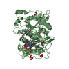
| Related structure data | 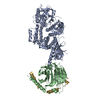 1omwS S: Starting model for refinement |
|---|---|
| Similar structure data |


-Links
 PDBj
PDBj


- Assembly
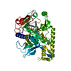
| Deposited unit | 
| ||||||||
|---|---|---|---|---|---|---|---|---|---|
| 1 |
| ||||||||
| Unit cell |
|
-Components
| #1: Protein | Mass: 37416.930 Da / Num. of mol.: 1 Source method: isolated from a genetically manipulated source Source: (gene. exp.)   Spodoptera frugiperda (fall armyworm) / Strain (production host): High 5 / References: UniProt: P62871 Spodoptera frugiperda (fall armyworm) / Strain (production host): High 5 / References: UniProt: P62871 |
|---|---|
| #2: Protein | Mass: 8690.024 Da / Num. of mol.: 1 Source method: isolated from a genetically manipulated source Source: (gene. exp.) Spodoptera frugiperda (fall armyworm) / Strain (production host): High5 / References: UniProt: P63212 |
| #3: Protein/peptide | Mass: 1688.919 Da / Num. of mol.: 1 / Source method: obtained synthetically Details: The peptide was chemically synthesized. This sequence is derived from a randomized phage library. |
| #4: Water | ChemComp-HOH /  Mass: 18.015 Da / Num. of mol.: 37 / Source method: isolated from a natural source / Formula: H2O Mass: 18.015 Da / Num. of mol.: 37 / Source method: isolated from a natural source / Formula: H2O |
-Experimental details
-Experiment
| Experiment | Method: X-RAY DIFFRACTION / Number of used crystals: 1 |
|---|
- Sample preparation
| Crystal | Density Matthews: 2.14 Å3/Da / Density % sol: 42.1 % |
|---|---|
| Crystal grow | Temperature: 293 K / Method: vapor diffusion, hanging drop / pH: 7.5 Details: PEG 4000, GLYCEROL, HEPES, PH 7.5, VAPOR DIFFUSION, HANGING DROP, temperature 293K |
-Data collection
| Diffraction | Mean temperature: 100 K |
|---|---|
| Diffraction source | Source: SYNCHROTRON / Site: ALS  / Beamline: 8.2.2 / Wavelength: 1.0871 Å / Beamline: 8.2.2 / Wavelength: 1.0871 Å |
| Detector | Type: ADSC QUANTUM 315 / Detector: CCD / Date: Nov 18, 2003 / Details: mirrors |
| Radiation | Monochromator: DOUBLE CRYSTAL SI (111) / Protocol: SINGLE WAVELENGTH / Monochromatic (M) / Laue (L): M / Scattering type: x-ray |
| Radiation wavelength | Wavelength: 1.0871 Å / Relative weight: 1 |
| Reflection | Resolution: 2.7→50 Å / Num. all: 9729 / Num. obs: 9729 / % possible obs: 88.3 % / Observed criterion σ(F): 0 / Observed criterion σ(I): -2.5 / Redundancy: 3.5 % / Biso Wilson estimate: 61.8 Å2 / Rsym value: 0.087 / Net I/σ(I): 13.5 |
| Reflection shell | Resolution: 2.7→2.8 Å / Redundancy: 1.8 % / Rmerge(I) obs: 0.414 / Mean I/σ(I) obs: 1.6 / Num. unique all: 408 / % possible all: 38.6 |
- Processing
| Software |
| ||||||||||||||||||||||||||||||||||||||||||||||||||||||||||||
|---|---|---|---|---|---|---|---|---|---|---|---|---|---|---|---|---|---|---|---|---|---|---|---|---|---|---|---|---|---|---|---|---|---|---|---|---|---|---|---|---|---|---|---|---|---|---|---|---|---|---|---|---|---|---|---|---|---|---|---|---|---|
| Refinement | Method to determine structure: MOLECULAR REPLACEMENT Starting model: PDB ENTRY 1OMW Resolution: 2.7→41.91 Å / Rfactor Rfree error: 0.01 / Data cutoff high absF: 2166288.6 / Data cutoff low absF: 0 / Isotropic thermal model: GROUP / Cross valid method: THROUGHOUT / σ(F): 0 / Stereochemistry target values: Engh & Huber
| ||||||||||||||||||||||||||||||||||||||||||||||||||||||||||||
| Solvent computation | Solvent model: FLAT MODEL / Bsol: 46.0853 Å2 / ksol: 0.346454 e/Å3 | ||||||||||||||||||||||||||||||||||||||||||||||||||||||||||||
| Displacement parameters | Biso mean: 46.3 Å2
| ||||||||||||||||||||||||||||||||||||||||||||||||||||||||||||
| Refine analyze |
| ||||||||||||||||||||||||||||||||||||||||||||||||||||||||||||
| Refinement step | Cycle: LAST / Resolution: 2.7→41.91 Å
| ||||||||||||||||||||||||||||||||||||||||||||||||||||||||||||
| Refine LS restraints |
| ||||||||||||||||||||||||||||||||||||||||||||||||||||||||||||
| LS refinement shell | Resolution: 2.7→2.87 Å / Rfactor Rfree error: 0.052 / Total num. of bins used: 6
| ||||||||||||||||||||||||||||||||||||||||||||||||||||||||||||
| Xplor file |
|
