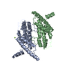
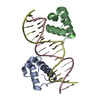
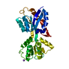
DNA BINDING PROTEIN / TELOMERE / DNA-BINDING PROTEIN / HOMEODOMAIN / MITOSIS / CELL CYCLE / NUCLEAR PROTEIN / CHROMOSOMAL PROTEIN / PHOSPHORYLATION / ADP-RIBOSYLATION
Function / homology
Function and homology information
axonal transport of messenger ribonucleoprotein complex / negative regulation of telomere single strand break repair / negative regulation of telomere maintenance via recombination / telomeric loop formation / negative regulation of telomere capping / protection from non-homologous end joining at telomere / negative regulation of telomere maintenance via semi-conservative replication / negative regulation of telomeric D-loop disassembly / RNA-templated DNA biosynthetic process / negative regulation of telomere maintenance ...axonal transport of messenger ribonucleoprotein complex / negative regulation of telomere single strand break repair / negative regulation of telomere maintenance via recombination / telomeric loop formation / negative regulation of telomere capping / protection from non-homologous end joining at telomere / negative regulation of telomere maintenance via semi-conservative replication / negative regulation of telomeric D-loop disassembly / RNA-templated DNA biosynthetic process / negative regulation of telomere maintenance / negative regulation of t-circle formation / regulation of telomere maintenance via telomerase / shelterin complex / telomeric D-loop disassembly / Telomere C-strand synthesis initiation / double-stranded telomeric DNA binding / Telomere C-strand (Lagging Strand) Synthesis / nuclear telomere cap complex / G-rich strand telomeric DNA binding / telomere capping / Polymerase switching on the C-strand of the telomere / Processive synthesis on the C-strand of the telomere / Removal of the Flap Intermediate from the C-strand / regulation of telomere maintenance / protein localization to chromosome, telomeric region / negative regulation of telomere maintenance via telomere lengthening / telomeric repeat DNA binding / negative regulation of telomere maintenance via telomerase / positive regulation of telomere maintenance / negative regulation of cellular senescence / Telomere Extension By Telomerase / Packaging Of Telomere Ends / Recognition and association of DNA glycosylase with site containing an affected purine / Cleavage of the damaged purine / Recognition and association of DNA glycosylase with site containing an affected pyrimidine / Cleavage of the damaged pyrimidine / telomere maintenance / Inhibition of DNA recombination at telomere / Meiotic synapsis / male germ cell nucleus / DNA Damage/Telomere Stress Induced Senescence / cellular senescence / in utero embryonic development / chromosome, telomeric region / nuclear body / axon / protein-containing complex binding / enzyme binding / protein homodimerization activity / nucleoplasm / nucleus Similarity search - Function
In the structure databanks used in Yorodumi, some data are registered as the other names, "COVID-19 virus" and "2019-nCoV". Here are the details of the virus and the list of structure data.
Jan 31, 2019. EMDB accession codes are about to change! (news from PDBe EMDB page)
EMDB accession codes are about to change! (news from PDBe EMDB page)
The allocation of 4 digits for EMDB accession codes will soon come to an end. Whilst these codes will remain in use, new EMDB accession codes will include an additional digit and will expand incrementally as the available range of codes is exhausted. The current 4-digit format prefixed with “EMD-” (i.e. EMD-XXXX) will advance to a 5-digit format (i.e. EMD-XXXXX), and so on. It is currently estimated that the 4-digit codes will be depleted around Spring 2019, at which point the 5-digit format will come into force.
The EM Navigator/Yorodumi systems omit the EMD- prefix.
Related info.:Q: What is EMD? / ID/Accession-code notation in Yorodumi/EM Navigator
Yorodumi is a browser for structure data from EMDB, PDB, SASBDB, etc.
This page is also the successor to EM Navigator detail page, and also detail information page/front-end page for Omokage search.
The word "yorodu" (or yorozu) is an old Japanese word meaning "ten thousand". "mi" (miru) is to see.
Related info.:EMDB / PDB / SASBDB / Comparison of 3 databanks / Yorodumi Search / Aug 31, 2016. New EM Navigator & Yorodumi / Yorodumi Papers / Jmol/JSmol / Function and homology information / Changes in new EM Navigator and Yorodumi
 Movie
Movie Controller


 Open data
 Basic information
 Components
 Keywords
 Function and homology information
 HOMO SAPIENS (human)
 X-RAY DIFFRACTION /
  Authors
 Citation
 Structure visualization
 Downloads & links
 Other downloads
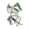
 PDBj
PDBj


 Assembly

 Mass: 18.015 Da / Num. of mol.: 130 / Source method: isolated from a natural source / Formula: H2O
 Sample preparation
 / Beamline: ID14-1 / Wavelength: 0.934
  Processing