 Movie
Movie Controller

[English] 日本語
 Yorodumi
- PDB-6nio: Crystal Structure of the Molybdate Transporter Periplasmic Protei... -
+ Open data

- Basic information
| Entry | Database: PDB / ID: 6nio | ||||||
|---|---|---|---|---|---|---|---|
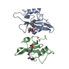
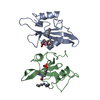
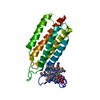
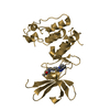
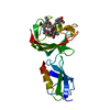
| Title | Crystal Structure of the Molybdate Transporter Periplasmic Protein ModA from Yersinia pestis | ||||||
 Components Components | Molybdate ABC transporter substrate-binding protein | ||||||
 Keywords Keywords | TRANSPORT PROTEIN / Periplasmic Protein ModA / Structural Genomics / Center for Structural Genomics of Infectious Diseases / CSGID | ||||||
| Function / homology | Periplasmic binding protein-like II / D-Maltodextrin-Binding Protein; domain 2 / 3-Layer(aba) Sandwich / Alpha Beta / ACETIC ACID / FORMIC ACID / :  Function and homology information Function and homology information | ||||||
| Biological species |   Yersinia pestis (bacteria) Yersinia pestis (bacteria) | ||||||
| Method |  X-RAY DIFFRACTION / SYNCHROTRON / SAD / Resolution: 1.37 Å X-RAY DIFFRACTION / SYNCHROTRON / SAD / Resolution: 1.37 Å | ||||||
 Authors Authors | Kim, Y. / Joachimiak, G. / Maltseva, N. / Endres, M. / Joachimiak, A. / Center for Structural Genomics of Infectious Diseases (CSGID) | ||||||
| Funding support |  United States, 1items United States, 1items
| ||||||
 Citation Citation | Journal: To Be Published Title: Crystal Structure of the Molybdate Transporter Periplasmic Protein ModA from Yersinia pestis Authors: Kim, Y. / Joachimiak, G. / Maltseva, N. / Endres, M. / Joachimiak, A. / Center for Structural Genomics of Infectious Diseases (CSGID) | ||||||
| History |
|
- Structure visualization
| Structure viewer | Molecule: MolmilJmol/JSmol |
|---|
- Downloads & links
-Download
| PDBx/mmCIF format | 6nio.cif.gz | 112.4 KB | Display | PDBx/mmCIF format |
|---|---|---|---|---|
| PDB format | pdb6nio.ent.gz | 84.3 KB | Display | PDB format |
| PDBx/mmJSON format | 6nio.json.gz | Tree view | PDBx/mmJSON format | |
| Others |  Other downloads Other downloads |
-Validation report
| Arichive directory | https://data.pdbj.org/pub/pdb/validation_reports/ni/6nioftp://data.pdbj.org/pub/pdb/validation_reports/ni/6nio | HTTPS FTP |
|---|
-Related structure data
| Similar structure data | |
|---|---|
| Other databases |


-Links
 PDBj
PDBj

- Assembly
| Deposited unit | 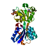
| ||||||||
|---|---|---|---|---|---|---|---|---|---|
| 1 |
| ||||||||
| Unit cell |
|
-Components
| #1: Protein | Mass: 25714.482 Da / Num. of mol.: 1 Source method: isolated from a genetically manipulated source Source: (gene. exp.) Yersinia pestis (bacteria) / Gene: modA, modA2, YP_1015, C6P89_14820, NCTC144_03853 / Plasmid: pMCSG68 / Production host: |
|---|---|
| #2: Chemical | ChemComp-NHE /   Mass: 207.290 Da / Num. of mol.: 1 / Source method: obtained synthetically / Formula: C8H17NO3S / Comment: pH buffer*YM Mass: 207.290 Da / Num. of mol.: 1 / Source method: obtained synthetically / Formula: C8H17NO3S / Comment: pH buffer*YM |
| #3: Chemical | ChemComp-ACY /   Mass: 60.052 Da / Num. of mol.: 1 / Source method: obtained synthetically / Formula: C2H4O2 Mass: 60.052 Da / Num. of mol.: 1 / Source method: obtained synthetically / Formula: C2H4O2 |
| #4: Chemical | ChemComp-FMT /   Mass: 46.025 Da / Num. of mol.: 1 / Source method: obtained synthetically / Formula: CH2O2 Mass: 46.025 Da / Num. of mol.: 1 / Source method: obtained synthetically / Formula: CH2O2 |
| #5: Water | ChemComp-HOH /  Mass: 18.015 Da / Num. of mol.: 191 / Source method: isolated from a natural source / Formula: H2O Mass: 18.015 Da / Num. of mol.: 191 / Source method: isolated from a natural source / Formula: H2O |
| Has protein modification | Y |
-Experimental details
-Experiment
| Experiment | Method: X-RAY DIFFRACTION / Number of used crystals: 1 |
|---|
- Sample preparation
| Crystal | Density Matthews: 1.93 Å3/Da / Density % sol: 36.17 % |
|---|---|
| Crystal grow | Temperature: 289 K / Method: vapor diffusion, sitting drop / pH: 9.5 / Details: 0.1 M CHES pH 9.5, 1.0 M sodium citrate |
-Data collection
| Diffraction | Mean temperature: 100 K / Serial crystal experiment: N |
|---|---|
| Diffraction source | Source: SYNCHROTRON / Site: APS / Beamline: 19-ID / Wavelength: 0.97927 Å |
| Detector | Type: DECTRIS PILATUS3 X 6M / Detector: PIXEL / Date: Nov 20, 2018 |
| Radiation | Protocol: SINGLE WAVELENGTH / Monochromatic (M) / Laue (L): M / Scattering type: x-ray |
| Radiation wavelength | Wavelength: 0.97927 Å / Relative weight: 1 |
| Reflection | Resolution: 1.37→50 Å / Num. obs: 41969 / % possible obs: 99.8 % / Redundancy: 11.9 % / Biso Wilson estimate: 19.01 Å2 / Rmerge(I) obs: 0.12 / Net I/σ(I): 35.9 |
| Reflection shell | Resolution: 1.37→1.39 Å / Redundancy: 11.6 % / Rmerge(I) obs: 0.687 / Mean I/σ(I) obs: 3.25 / Num. unique obs: 2078 / CC1/2: 0.883 / % possible all: 99.8 |
- Processing
| Software |
| ||||||||||||||||||||||||||||||||||||||||||||||||||||||||||||||||||||||||||||||||||||||||||||||||||||||||||||||||
|---|---|---|---|---|---|---|---|---|---|---|---|---|---|---|---|---|---|---|---|---|---|---|---|---|---|---|---|---|---|---|---|---|---|---|---|---|---|---|---|---|---|---|---|---|---|---|---|---|---|---|---|---|---|---|---|---|---|---|---|---|---|---|---|---|---|---|---|---|---|---|---|---|---|---|---|---|---|---|---|---|---|---|---|---|---|---|---|---|---|---|---|---|---|---|---|---|---|---|---|---|---|---|---|---|---|---|---|---|---|---|---|---|---|
| Refinement | Method to determine structure: SAD / Resolution: 1.37→41.657 Å / SU ML: 0.11 / Cross valid method: FREE R-VALUE / σ(F): 1.39 / Phase error: 17.39
| ||||||||||||||||||||||||||||||||||||||||||||||||||||||||||||||||||||||||||||||||||||||||||||||||||||||||||||||||
| Solvent computation | Shrinkage radii: 0.9 Å / VDW probe radii: 1.11 Å | ||||||||||||||||||||||||||||||||||||||||||||||||||||||||||||||||||||||||||||||||||||||||||||||||||||||||||||||||
| Displacement parameters | Biso mean: 24.6 Å2 | ||||||||||||||||||||||||||||||||||||||||||||||||||||||||||||||||||||||||||||||||||||||||||||||||||||||||||||||||
| Refinement step | Cycle: LAST / Resolution: 1.37→41.657 Å
| ||||||||||||||||||||||||||||||||||||||||||||||||||||||||||||||||||||||||||||||||||||||||||||||||||||||||||||||||
| Refine LS restraints |
| ||||||||||||||||||||||||||||||||||||||||||||||||||||||||||||||||||||||||||||||||||||||||||||||||||||||||||||||||
| LS refinement shell |
|
