 Movie
Movie Controller

[English] 日本語
 Yorodumi
- PDB-1v1i: Adenovirus fibre shaft sequence N-terminally fused to the bacteri... -
+ Open data

- Basic information
| Entry | Database: PDB / ID: 1v1i | ||||||
|---|---|---|---|---|---|---|---|
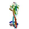
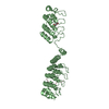
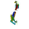
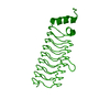
| Title | Adenovirus fibre shaft sequence N-terminally fused to the bacteriophage T4 fibritin foldon trimerisation motif with a long linker | ||||||
 Components Components | FIBRITIN, FIBER PROTEIN | ||||||
 Keywords Keywords | ADENOVIRUS / CHIMERA / FIBER PROTEIN | ||||||
| Function / homology |  Function and homology information Function and homology informationadhesion receptor-mediated virion attachment to host cell / virion component / viral capsid / cell adhesion / symbiont entry into host cell / host cell nucleus Similarity search - Function | ||||||
| Biological species |  HUMAN ADENOVIRUS CENTEROBACTERIA PHAGE T4 (virus) HUMAN ADENOVIRUS CENTEROBACTERIA PHAGE T4 (virus) | ||||||
| Method |  X-RAY DIFFRACTION / SYNCHROTRON / MOLECULAR REPLACEMENT / Resolution: 1.9 Å X-RAY DIFFRACTION / SYNCHROTRON / MOLECULAR REPLACEMENT / Resolution: 1.9 Å | ||||||
 Authors Authors | Papanikolopoulou, K. / Teixeira, S. / Belrhali, H. / Forsyth, V.T. / Mitraki, A. / van Raaij, M.J. | ||||||
 Citation Citation | Journal: J.Mol.Biol. / Year: 2004 Title: Adenovirus Fibre Shaft Sequences Fold Into the Native Triple Beta-Spiral Fold When N-Terminally Fused to the Bacteriophage T4 Fibritin Foldon Trimerisation Motif Authors: Papanikolopoulou, K. / Teixeira, S. / Belrhali, H. / Forsyth, V.T. / Mitraki, A. / van Raaij, M.J. #1: Journal: J.Biol.Chem. / Year: 2004 Title: Formation of Highly Stable Chimeric Trimers by Fusion of an Adenovirus Fiber Shaft Fragment with the Foldon Domain of Bacteriophage T4 Fibritin Authors: Papanikolopoulou, K. / Forge, V. / Goeltz, P. / Mitraki, A. #2: Journal: Nature / Year: 1999Title: A Triple Beta-Spiral in the Adenovirus Fibre Shaft Reveals a New Structural Motif for a Fibrous Protein Authors: van Raaij, M.J. / Mitraki, A. / Lavigne, G. / Cusack, S. #3: Journal: Acta Crystallogr.,Sect.D / Year: 1998Title: Structure of Bacteriophage T4 Fibritin M: A Troublesome Packing Arrangement Authors: Strelkov, S. / Tao, Y. / Shneider, M.M. / Mesyanzhinov, V. / Rossmann, M.G. | ||||||
| History |
|
- Structure visualization
| Structure viewer | Molecule: MolmilJmol/JSmol |
|---|
- Downloads & links
-Download
| PDBx/mmCIF format | 1v1i.cif.gz | 75.1 KB | Display | PDBx/mmCIF format |
|---|---|---|---|---|
| PDB format | pdb1v1i.ent.gz | 57.1 KB | Display | PDB format |
| PDBx/mmJSON format | 1v1i.json.gz | Tree view | PDBx/mmJSON format | |
| Others |  Other downloads Other downloads |
-Validation report
| Arichive directory | https://data.pdbj.org/pub/pdb/validation_reports/v1/1v1iftp://data.pdbj.org/pub/pdb/validation_reports/v1/1v1i | HTTPS FTP |
|---|
-Related structure data
| Related structure data |  1v1hC  1avyS  1qiuS S: Starting model for refinement C: citing same article ( |
|---|---|
| Similar structure data |


-Links
 PDBj
PDBj


- Assembly
| Deposited unit | 
| ||||||||||||||||||||||||||||
|---|---|---|---|---|---|---|---|---|---|---|---|---|---|---|---|---|---|---|---|---|---|---|---|---|---|---|---|---|---|
| 1 |
| ||||||||||||||||||||||||||||
| Unit cell |
| ||||||||||||||||||||||||||||
| Noncrystallographic symmetry (NCS) | NCS oper:
|
-Components
| #1: Protein | Mass: 11509.702 Da / Num. of mol.: 3 Fragment: SHAFT DOMAIN PLUS FOLDON DOMAIN, RESIDUES 319-392 AND 457-483 Source method: isolated from a genetically manipulated source Details: ARTIFICIAL FUSION PROTEIN OF ADENOVIRUS TYPE 2 FIBRE SHAFT RESIDUES 319-398 - BACTERIOPHAGE T4 FIBRITIN FOLDON RESIDUES 457-483 WITH A GLY-SER LINKER IN BETWEEN Source: (gene. exp.) HUMAN ADENOVIRUS C, (gene. exp.) ENTEROBACTERIA PHAGE T4 (virus)Plasmid: PT7.7 / Production host:  #2: Water | ChemComp-HOH / |  Mass: 18.015 Da / Num. of mol.: 237 / Source method: isolated from a natural source / Formula: H2O Mass: 18.015 Da / Num. of mol.: 237 / Source method: isolated from a natural source / Formula: H2OCompound details | ADENOVIRUS FIBRE IS RESPONSIBLE FOR ADENOVIRUS RECEPTOR BINDING AND CONTAINS A VIRUS-BINDING N- ...ADENOVIRUS | |
|---|
-Experimental details
-Experiment
| Experiment | Method: X-RAY DIFFRACTION / Number of used crystals: 1 |
|---|
- Sample preparation
| Crystal | Density Matthews: 2.2 Å3/Da / Density % sol: 44.8 % |
|---|---|
| Crystal grow | pH: 7 Details: 10 MM HEPES-NAOH PH 7.0 0.2 M MAGNESIUM ACETATE, 20 % (W/V) PEG 3350 |
-Data collection
| Diffraction | Mean temperature: 100 K |
|---|---|
| Diffraction source | Source: SYNCHROTRON / Site: ESRF  / Beamline: BM14 / Wavelength: 0.9202 / Beamline: BM14 / Wavelength: 0.9202 |
| Detector | Type: MARRESEARCH / Detector: CCD / Date: Sep 24, 2003 |
| Radiation | Protocol: SINGLE WAVELENGTH / Monochromatic (M) / Laue (L): M / Scattering type: x-ray |
| Radiation wavelength | Wavelength: 0.9202 Å / Relative weight: 1 |
| Reflection | Resolution: 1.9→20 Å / Num. obs: 25299 / % possible obs: 99.9 % / Redundancy: 13.8 % / Biso Wilson estimate: 25.453 Å2 / Rmerge(I) obs: 0.068 / Rsym value: 0.068 / Net I/σ(I): 6.9 |
| Reflection shell | Resolution: 1.9→2 Å / Redundancy: 10.5 % / Rmerge(I) obs: 0.33 / Mean I/σ(I) obs: 2.2 / Rsym value: 0.33 / % possible all: 99.8 |
- Processing
| Software |
| ||||||||||||||||||||
|---|---|---|---|---|---|---|---|---|---|---|---|---|---|---|---|---|---|---|---|---|---|
| Refinement | Method to determine structure: MOLECULAR REPLACEMENT Starting model: PDB ENTRY 1QIU, PDB ENTRY 1AVY Resolution: 1.9→19.9 Å / SU B: 4.5422 / SU ML: 0.1332 / Cross valid method: THROUGHOUT / ESU R: 0.1933 / ESU R Free: 0.1788
| ||||||||||||||||||||
| Displacement parameters | Biso mean: 32.746 Å2
| ||||||||||||||||||||
| Refinement step | Cycle: LAST / Resolution: 1.9→19.9 Å
|
