 Movie
Movie Controller

[English] 日本語
 Yorodumi
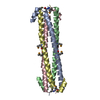
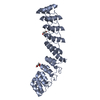
- PDB-1v1h: Adenovirus fibre shaft sequence N-terminally fused to the bacteri... -
+ Open data

- Basic information
| Entry | Database: PDB / ID: 1v1h | ||||||
|---|---|---|---|---|---|---|---|
| Title | Adenovirus fibre shaft sequence N-terminally fused to the bacteriophage T4 fibritin foldon trimerisation motif with a short linker | ||||||
 Components Components | FIBRITIN, FIBER PROTEIN | ||||||
 Keywords Keywords | ADENOVIRUS / CHIMERA / FIBER PROTEIN | ||||||
| Function / homology |  Function and homology information Function and homology informationadhesion receptor-mediated virion attachment to host cell / virion component / viral capsid / cell adhesion / symbiont entry into host cell / host cell nucleus Similarity search - Function | ||||||
| Biological species |   HUMAN ADENOVIRUS TYPE 2BACTERIOPHAGE T4 (virus) HUMAN ADENOVIRUS TYPE 2BACTERIOPHAGE T4 (virus) | ||||||
| Method |  X-RAY DIFFRACTION / SYNCHROTRON / MOLECULAR REPLACEMENT / Resolution: 1.9 Å X-RAY DIFFRACTION / SYNCHROTRON / MOLECULAR REPLACEMENT / Resolution: 1.9 Å | ||||||
 Authors Authors | Papanikolopoulou, K. / Teixeira, S. / Belrhali, H. / Forsyth, V.T. / Mitraki, A. / van Raaij, M.J. | ||||||
 Citation Citation | Journal: J.Mol.Biol. / Year: 2004 Title: Adenovirus Fibre Shaft Sequences Fold Into the Native Triple Beta-Spiral Fold When N-Terminally Fused to the Bacteriophage T4 Fibritin Foldon Trimerisation Motif Authors: Papanikolopoulou, K. / Teixeira, S. / Belrhali, H. / Forsyth, V.T. / Mitraki, A. / van Raaij, M.J. #1: Journal: J.Biol.Chem. / Year: 2004 Title: Formation of Highly Stable Chimeric Trimers by Fusion of an Adenovirus Fiber Shaft Fragment with the Foldon Domain of Bacteriophage T4 Fibritin Authors: Papanikolopoulou, K. / Forge, V. / Goeltz, P. / Mitraki, A. #2: Journal: Nature / Year: 1999Title: A Triple Beta-Spiral in the Adenovirus Fibre Shaft Reveals a New Structural Motif for a Fibrous Protein Authors: van Raaij, M.J. / Mitraki, A. / Lavigne, G. / Cusack, S. #3: Journal: Acta Crystallogr.,Sect.D / Year: 1998Title: Structure of Bacteriophage T4 Fibritin M: A Troublesome Packing Arrangement Authors: Strelkov, S. / Tao, Y. / Shneider, M.M. / Mesyanzhinov, V. / Rossmann, M.G. | ||||||
| History |
| ||||||
| Remark 700 | SHEET THE SHEET STRUCTURE OF THIS MOLECULE IS BIFURCATED. IN ORDER TO REPRESENT THIS FEATURE IN ... SHEET THE SHEET STRUCTURE OF THIS MOLECULE IS BIFURCATED. IN ORDER TO REPRESENT THIS FEATURE IN THE SHEET RECORDS BELOW, TWO SHEETS ARE DEFINED. |
- Structure visualization
| Structure viewer | Molecule: MolmilJmol/JSmol |
|---|
- Downloads & links
-Download
| PDBx/mmCIF format | 1v1h.cif.gz | 135.3 KB | Display | PDBx/mmCIF format |
|---|---|---|---|---|
| PDB format | pdb1v1h.ent.gz | 108.9 KB | Display | PDB format |
| PDBx/mmJSON format | 1v1h.json.gz | Tree view | PDBx/mmJSON format | |
| Others |  Other downloads Other downloads |
-Validation report
| Arichive directory | https://data.pdbj.org/pub/pdb/validation_reports/v1/1v1hftp://data.pdbj.org/pub/pdb/validation_reports/v1/1v1h | HTTPS FTP |
|---|
-Related structure data
| Related structure data |  1v1iC  1qiuS S: Starting model for refinement C: citing same article ( |
|---|---|
| Similar structure data |


-Links
 PDBj
PDBj


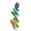
- Assembly
| Deposited unit | 
| ||||||||||||||||||||||||||||||||||||||||||||||||||||||||||||||||||||||||||||||||||||||||||||||||||||||||||||||||||
|---|---|---|---|---|---|---|---|---|---|---|---|---|---|---|---|---|---|---|---|---|---|---|---|---|---|---|---|---|---|---|---|---|---|---|---|---|---|---|---|---|---|---|---|---|---|---|---|---|---|---|---|---|---|---|---|---|---|---|---|---|---|---|---|---|---|---|---|---|---|---|---|---|---|---|---|---|---|---|---|---|---|---|---|---|---|---|---|---|---|---|---|---|---|---|---|---|---|---|---|---|---|---|---|---|---|---|---|---|---|---|---|---|---|---|---|
| 1 | 
| ||||||||||||||||||||||||||||||||||||||||||||||||||||||||||||||||||||||||||||||||||||||||||||||||||||||||||||||||||
| 2 | 
| ||||||||||||||||||||||||||||||||||||||||||||||||||||||||||||||||||||||||||||||||||||||||||||||||||||||||||||||||||
| Unit cell |
| ||||||||||||||||||||||||||||||||||||||||||||||||||||||||||||||||||||||||||||||||||||||||||||||||||||||||||||||||||
| Components on special symmetry positions |
| ||||||||||||||||||||||||||||||||||||||||||||||||||||||||||||||||||||||||||||||||||||||||||||||||||||||||||||||||||
| Noncrystallographic symmetry (NCS) | NCS oper:
|
