 Movie
Movie Controller

[English] 日本語
 Yorodumi
- PDB-1qiu: A triple beta-spiral in the adenovirus fibre shaft reveals a new ... -
+ Open data

- Basic information
| Entry | Database: PDB / ID: 1qiu | ||||||
|---|---|---|---|---|---|---|---|
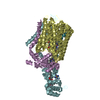
| Title | A triple beta-spiral in the adenovirus fibre shaft reveals a new structural motif for biological fibres | ||||||
 Components Components | ADENOVIRUS FIBRE | ||||||
 Keywords Keywords | FIBRE PROTEIN / TRIPLE BETA-SPIRAL / ADENOVIRUS | ||||||
| Function / homology |  Function and homology information Function and homology informationadhesion receptor-mediated virion attachment to host cell / viral capsid / cell adhesion / symbiont entry into host cell / host cell nucleus Similarity search - Function | ||||||
| Biological species |   HUMAN ADENOVIRUS 2 HUMAN ADENOVIRUS 2 | ||||||
| Method |  X-RAY DIFFRACTION / SYNCHROTRON / MOLECULAR REPLACEMENT / Resolution: 2.4 Å X-RAY DIFFRACTION / SYNCHROTRON / MOLECULAR REPLACEMENT / Resolution: 2.4 Å | ||||||
 Authors Authors | van Raaij, M.J. / Lavigne, G. / Mitraki, A. / Cusack, S. | ||||||
 Citation Citation | Journal: Nature / Year: 1999 Title: A triple beta-spiral in the adenovirus fibre shaft reveals a new structural motif for a fibrous protein. Authors: van Raaij, M.J. / Mitraki, A. / Lavigne, G. / Cusack, S. | ||||||
| History |
|
- Structure visualization
| Structure viewer | Molecule: MolmilJmol/JSmol |
|---|
- Downloads & links
-Download
| PDBx/mmCIF format | 1qiu.cif.gz | 311 KB | Display | PDBx/mmCIF format |
|---|---|---|---|---|
| PDB format | pdb1qiu.ent.gz | 255.6 KB | Display | PDB format |
| PDBx/mmJSON format | 1qiu.json.gz | Tree view | PDBx/mmJSON format | |
| Others |  Other downloads Other downloads |
-Validation report
| Arichive directory | https://data.pdbj.org/pub/pdb/validation_reports/qi/1qiuftp://data.pdbj.org/pub/pdb/validation_reports/qi/1qiu | HTTPS FTP |
|---|
-Related structure data
| Related structure data | 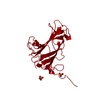 1qhvS S: Starting model for refinement |
|---|---|
| Similar structure data |


-Links
 PDBj
PDBj
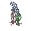
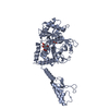
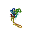
- Assembly
| Deposited unit | 
| ||||||||
|---|---|---|---|---|---|---|---|---|---|
| 1 | 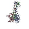
| ||||||||
| 2 | 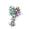
| ||||||||
| Unit cell |
| ||||||||
| Details | THE MOLECULE CONSISTS IS A HOMO- TRIMER.CHAINS A, B AND C FORM ONE TRIMER CHAINS D, E AND F THE OTHER TRIMER |
-Components
| #1: Protein | Mass: 28587.725 Da / Num. of mol.: 6 Fragment: FRAGMENT CONTAINING THE FOUR DISTAL SHAFT REPEATS PLUS THE HEAD DOMAIN. Source method: isolated from a genetically manipulated source Source: (gene. exp.) HUMAN ADENOVIRUS 2 / Gene: LOCUS AD2H2 / Plasmid: PT7.7 / Cellular location (production host): CYTOPLASM / Production host:  #2: Water | ChemComp-HOH / |  Mass: 18.015 Da / Num. of mol.: 579 / Source method: isolated from a natural source / Formula: H2O Mass: 18.015 Da / Num. of mol.: 579 / Source method: isolated from a natural source / Formula: H2O |
|---|
-Experimental details
-Experiment
| Experiment | Method: X-RAY DIFFRACTION / Number of used crystals: 1 |
|---|
- Sample preparation
| Crystal | Density Matthews: 4.6 Å3/Da / Density % sol: 73 % | ||||||||||||||||||||
|---|---|---|---|---|---|---|---|---|---|---|---|---|---|---|---|---|---|---|---|---|---|
| Crystal grow | pH: 4 Details: 0.8 M AMMONIUM SULPHATE, 100 MM SODIUM CITRATE, PH 4.0 | ||||||||||||||||||||
| Crystal | *PLUS | ||||||||||||||||||||
| Crystal grow | *PLUS Temperature: 20 ℃ / Method: vapor diffusion, sitting drop | ||||||||||||||||||||
| Components of the solutions | *PLUS
|
-Data collection
| Diffraction | Mean temperature: 100 K |
|---|---|
| Diffraction source | Source: SYNCHROTRON / Site: ESRF  / Beamline: ID14-3 / Wavelength: 0.948 / Beamline: ID14-3 / Wavelength: 0.948 |
| Detector | Type: MARRESEARCH / Detector: CCD / Date: Sep 5, 1998 |
| Radiation | Protocol: SINGLE WAVELENGTH / Monochromatic (M) / Laue (L): M / Scattering type: x-ray |
| Radiation wavelength | Wavelength: 0.948 Å / Relative weight: 1 |
| Reflection | Resolution: 2.4→25 Å / Num. obs: 103327 / % possible obs: 84 % / Observed criterion σ(I): 0 / Redundancy: 2.7 % / Biso Wilson estimate: 35.3 Å2 / Rsym value: 0.132 / Net I/σ(I): 4.1 |
| Reflection shell | Resolution: 2.4→2.53 Å / Redundancy: 1.7 % / Mean I/σ(I) obs: 2.4 / Rsym value: 0.287 / % possible all: 54.9 |
| Reflection | *PLUS Rmerge(I) obs: 0.132 |
| Reflection shell | *PLUS % possible obs: 59.9 % / Num. unique obs: 10696 / Rmerge(I) obs: 0.287 |
- Processing
| Software |
| ||||||||||||||||||||||||||||||||||||||||||||||||||||||||||||
|---|---|---|---|---|---|---|---|---|---|---|---|---|---|---|---|---|---|---|---|---|---|---|---|---|---|---|---|---|---|---|---|---|---|---|---|---|---|---|---|---|---|---|---|---|---|---|---|---|---|---|---|---|---|---|---|---|---|---|---|---|---|
| Refinement | Method to determine structure: MOLECULAR REPLACEMENT Starting model: PDB ENTRY 1QHV Resolution: 2.4→25 Å / Rfactor Rfree error: 0.006 / Data cutoff high absF: 2480815.2 / Isotropic thermal model: RESTRAINED / Cross valid method: THROUGHOUT / σ(F): 0 Stereochemistry target values: MAXIMUM LIKELIHOOD TARGET USING AMPLITUDES
| ||||||||||||||||||||||||||||||||||||||||||||||||||||||||||||
| Solvent computation | Solvent model: FLAT MODEL / Bsol: 44 Å2 / ksol: 0.38 e/Å3 | ||||||||||||||||||||||||||||||||||||||||||||||||||||||||||||
| Displacement parameters | Biso mean: 35.8 Å2
| ||||||||||||||||||||||||||||||||||||||||||||||||||||||||||||
| Refine analyze |
| ||||||||||||||||||||||||||||||||||||||||||||||||||||||||||||
| Refinement step | Cycle: LAST / Resolution: 2.4→25 Å
| ||||||||||||||||||||||||||||||||||||||||||||||||||||||||||||
| Refine LS restraints |
| ||||||||||||||||||||||||||||||||||||||||||||||||||||||||||||
| LS refinement shell | Resolution: 2.4→2.53 Å / Rfactor Rfree error: 0.025 / Total num. of bins used: 7
| ||||||||||||||||||||||||||||||||||||||||||||||||||||||||||||
| Xplor file |
| ||||||||||||||||||||||||||||||||||||||||||||||||||||||||||||
| Software | *PLUS Name: CNS / Version: 0.4 / Classification: refinement | ||||||||||||||||||||||||||||||||||||||||||||||||||||||||||||
| Refine LS restraints | *PLUS
| ||||||||||||||||||||||||||||||||||||||||||||||||||||||||||||
| LS refinement shell | *PLUS Rfactor obs: 0.312 |
