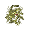
 Movie
Movie Controller


+ Open data

- Basic information
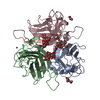
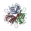
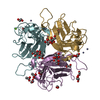
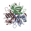
| Entry | Database: PDB / ID: 1qhv | ||||||
|---|---|---|---|---|---|---|---|
| Title | HUMAN ADENOVIRUS SEROTYPE 2 FIBRE HEAD | ||||||
 Components Components | PROTEIN (ADENOVIRUS FIBRE) | ||||||
 Keywords Keywords | VIRAL PROTEIN / RECEPTOR BINDING / EXTRA-ORDINARY STABILITY | ||||||
| Function / homology |  Function and homology information Function and homology informationadhesion receptor-mediated virion attachment to host cell / viral capsid / cell adhesion / symbiont entry into host cell / host cell nucleus Similarity search - Function | ||||||
| Biological species |   Human adenovirus 2 Human adenovirus 2 | ||||||
| Method |  X-RAY DIFFRACTION / SYNCHROTRON / MOLECULAR REPLACEMENT / Resolution: 1.51 Å X-RAY DIFFRACTION / SYNCHROTRON / MOLECULAR REPLACEMENT / Resolution: 1.51 Å | ||||||
 Authors Authors | Van Raaij, M.J. / Louis, N. / Chroboczek, J. / Cusack, S. | ||||||
 Citation Citation | Journal: Virology / Year: 1999 Title: Structure of the human adenovirus serotype 2 fiber head domain at 1.5 A resolution. Authors: van Raaij, M.J. / Louis, N. / Chroboczek, J. / Cusack, S. #1: Journal: J.Virol. / Year: 1994Title: Cell-Binding Domain of Adenovirus Serotype 2 Fiber Authors: Louis, N. / Fender, P. / Barge, A. / Kitts, P. / Chroboczek, J. | ||||||
| History |
|
- Structure visualization
| Structure viewer | Molecule: MolmilJmol/JSmol |
|---|
- Downloads & links
-Download
| PDBx/mmCIF format | 1qhv.cif.gz | 102.5 KB | Display | PDBx/mmCIF format |
|---|---|---|---|---|
| PDB format | pdb1qhv.ent.gz | 78.3 KB | Display | PDB format |
| PDBx/mmJSON format | 1qhv.json.gz | Tree view | PDBx/mmJSON format | |
| Others |  Other downloads Other downloads |
-Validation report
| Arichive directory | https://data.pdbj.org/pub/pdb/validation_reports/qh/1qhvftp://data.pdbj.org/pub/pdb/validation_reports/qh/1qhv | HTTPS FTP |
|---|
-Related structure data
| Related structure data |  1knbS S: Starting model for refinement |
|---|---|
| Similar structure data |


-Links
 PDBj
PDBj
- Assembly
| Deposited unit | 
| ||||||||||||||||||
|---|---|---|---|---|---|---|---|---|---|---|---|---|---|---|---|---|---|---|---|
| 1 | 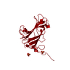
| ||||||||||||||||||
| Unit cell |
| ||||||||||||||||||
| Components on special symmetry positions |
| ||||||||||||||||||
| Details | THE FUNCTIONAL MOLECULE IS A TRIMER. THIS TRIMER IS GENERATED USING THE THREE-FOLD AXIS SYMMETRY. |
-Components
| #1: Protein | Mass: 21478.893 Da / Num. of mol.: 1 / Fragment: HEAD DOMAIN Source method: isolated from a genetically manipulated source Source: (gene. exp.) Human adenovirus 2 / Genus: Mastadenovirus / Species: Human adenovirus C / Strain: SEROTYPE 2 / Gene: LOCUS AD2H2 / Plasmid: PACCL29 / Production host:   Spodoptera frugiperda (fall armyworm) / Strain (production host): SF9 / References: UniProt: Q96590, UniProt: P03275*PLUS Spodoptera frugiperda (fall armyworm) / Strain (production host): SF9 / References: UniProt: Q96590, UniProt: P03275*PLUS | ||
|---|---|---|---|
| #2: Chemical |   Mass: 96.063 Da / Num. of mol.: 2 / Source method: obtained synthetically / Formula: SO4 Mass: 96.063 Da / Num. of mol.: 2 / Source method: obtained synthetically / Formula: SO4#3: Water | ChemComp-HOH / |  Mass: 18.015 Da / Num. of mol.: 305 / Source method: isolated from a natural source / Formula: H2O Mass: 18.015 Da / Num. of mol.: 305 / Source method: isolated from a natural source / Formula: H2O |
-Experimental details
-Experiment
| Experiment | Method: X-RAY DIFFRACTION / Number of used crystals: 3 |
|---|
- Sample preparation
| Crystal | Density Matthews: 2.96 Å3/Da / Density % sol: 58 % | |||||||||||||||||||||||||||||||||||
|---|---|---|---|---|---|---|---|---|---|---|---|---|---|---|---|---|---|---|---|---|---|---|---|---|---|---|---|---|---|---|---|---|---|---|---|---|
| Crystal grow | pH: 4 Details: 100 MM SODIUM ACETATE 25 % (V/V) GLYCEROL 1-1.5 M AMMONIUM SULPHATE PH 4.0 WITH ACETIC ACID | |||||||||||||||||||||||||||||||||||
| Crystal | *PLUS | |||||||||||||||||||||||||||||||||||
| Crystal grow | *PLUS Temperature: 20 ℃ / Method: vapor diffusion, sitting drop / PH range low: 5.2 / PH range high: 4 | |||||||||||||||||||||||||||||||||||
| Components of the solutions | *PLUS
|
-Data collection
| Diffraction | Mean temperature: 100 K |
|---|---|
| Diffraction source | Source: SYNCHROTRON / Site: ESRF  / Beamline: ID2 / Wavelength: 0.993 / Beamline: ID2 / Wavelength: 0.993 |
| Detector | Type: MARRESEARCH / Detector: IMAGE PLATE / Date: Apr 1, 1997 |
| Radiation | Protocol: SINGLE WAVELENGTH / Monochromatic (M) / Laue (L): M / Scattering type: x-ray |
| Radiation wavelength | Wavelength: 0.993 Å / Relative weight: 1 |
| Reflection | Resolution: 1.51→10.97 Å / Num. obs: 36820 / % possible obs: 91.8 % / Redundancy: 4.7 % / Biso Wilson estimate: 13.2 Å2 / Rsym value: 0.073 / Net I/σ(I): 7.3 |
| Reflection shell | Resolution: 1.51→1.59 Å / Redundancy: 1.4 % / Mean I/σ(I) obs: 3.6 / Rsym value: 0.172 / % possible all: 55.3 |
| Reflection | *PLUS Num. measured all: 386827 / Rmerge(I) obs: 0.073 |
| Reflection shell | *PLUS % possible obs: 55.3 % / Num. unique obs: 3170 / Rmerge(I) obs: 0.172 |
- Processing
| Software |
| ||||||||||||||||||||||||||||||||||||||||||||||||||||||||||||||||||||||||||||
|---|---|---|---|---|---|---|---|---|---|---|---|---|---|---|---|---|---|---|---|---|---|---|---|---|---|---|---|---|---|---|---|---|---|---|---|---|---|---|---|---|---|---|---|---|---|---|---|---|---|---|---|---|---|---|---|---|---|---|---|---|---|---|---|---|---|---|---|---|---|---|---|---|---|---|---|---|---|
| Refinement | Method to determine structure: MOLECULAR REPLACEMENT Starting model: PDB ENTRY 1KNB Resolution: 1.51→10.97 Å / SU B: 0.80142 / SU ML: 0.02897 / Cross valid method: THROUGHOUT / σ(F): 0 / ESU R: 0.05292 / ESU R Free: 0.05087
| ||||||||||||||||||||||||||||||||||||||||||||||||||||||||||||||||||||||||||||
| Displacement parameters | Biso mean: 23.07 Å2 | ||||||||||||||||||||||||||||||||||||||||||||||||||||||||||||||||||||||||||||
| Refinement step | Cycle: LAST / Resolution: 1.51→10.97 Å
| ||||||||||||||||||||||||||||||||||||||||||||||||||||||||||||||||||||||||||||
| Refine LS restraints |
| ||||||||||||||||||||||||||||||||||||||||||||||||||||||||||||||||||||||||||||
| Software | *PLUS Name: REFMAC / Classification: refinement | ||||||||||||||||||||||||||||||||||||||||||||||||||||||||||||||||||||||||||||
| Refinement | *PLUS % reflection Rfree: 3 % / Rfactor obs: 0.109 | ||||||||||||||||||||||||||||||||||||||||||||||||||||||||||||||||||||||||||||
| Solvent computation | *PLUS | ||||||||||||||||||||||||||||||||||||||||||||||||||||||||||||||||||||||||||||
| Displacement parameters | *PLUS | ||||||||||||||||||||||||||||||||||||||||||||||||||||||||||||||||||||||||||||
| Refine LS restraints | *PLUS
| ||||||||||||||||||||||||||||||||||||||||||||||||||||||||||||||||||||||||||||
| LS refinement shell | *PLUS Rfactor Rfree: 0.18 / Rfactor obs: 0.136 |
