 Movie
Movie Controller


+ Open data

- Basic information
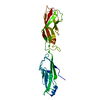
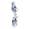
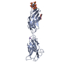
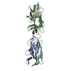
| Entry | Database: PDB / ID: 1avy | ||||||
|---|---|---|---|---|---|---|---|
| Title | FIBRITIN DELETION MUTANT M (BACTERIOPHAGE T4) | ||||||
 Components Components | FIBRITIN | ||||||
 Keywords Keywords | COILED COIL / BACTERIOPHAGE T4 / STRUCTURAL PROTEIN / CHAPERONE / BACTERIOPHAGE ASSEMBLY / PROTEIN FOLDING | ||||||
| Function / homology | 6-Phosphogluconate Dehydrogenase, domain 3 / Fibritin C-terminal / Fibritin C-terminal region / Single alpha-helices involved in coiled-coils or other helix-helix interfaces / virion component / Up-down Bundle / Mainly Alpha / Fibritin Function and homology information Function and homology information | ||||||
| Biological species |  Enterobacteria phage T4 (virus) Enterobacteria phage T4 (virus) | ||||||
| Method |  X-RAY DIFFRACTION / MOLECULAR REPLACEMENT / Resolution: 1.85 Å X-RAY DIFFRACTION / MOLECULAR REPLACEMENT / Resolution: 1.85 Å | ||||||
 Authors Authors | Strelkov, S.V. / Tao, Y. / Mesyanzhinov, V.V. / Rossmann, M.G. | ||||||
 Citation Citation | Journal: Acta Crystallogr.,Sect.D / Year: 1998 Title: Structure of bacteriophage T4 fibritin M: a troublesome packing arrangement. Authors: Strelkov, S.V. / Tao, Y. / Shneider, M.M. / Mesyanzhinov, V.V. / Rossmann, M.G. #1: Journal: Structure / Year: 1997Title: Structure of Bacteriophage T4 Fibritin: A Segmented Coiled Coil and the Role of the C-Terminal Domain Authors: Tao, Y. / Strelkov, S.V. / Mesyanzhinov, V.V. / Rossmann, M.G. #2: Journal: Virology / Year: 1996Title: Preliminary Crystallographic Studies of Bacteriophage T4 Fibritin Confirm a Trimeric Coiled-Coil Structure Authors: Strelkov, S.V. / Tao, Y. / Rossmann, M.G. / Kurochkina, L.P. / Shneider, M.M. / Mesyanzhinov, V.V. #3: Journal: J.Mol.Biol. / Year: 1994Title: Fibritin Encoded by Bacteriophage T4 Gene Wac Has a Parallel Triple-Stranded Alpha-Helical Coiled-Coil Structure Authors: Efimov, V.P. / Nepluev, I.V. / Sobolev, B.N. / Zurabishvili, T.G. / Schulthess, T. / Lustig, A. / Engel, J. / Haener, M. / Aebi, U. / Venyaminov, S.Yu. / Potekhin, S.A. / Mesyanzhinov, V.V. | ||||||
| History |
|
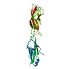
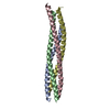
- Structure visualization
| Structure viewer | Molecule: MolmilJmol/JSmol |
|---|
- Downloads & links
-Download
| PDBx/mmCIF format | 1avy.cif.gz | 54.7 KB | Display | PDBx/mmCIF format |
|---|---|---|---|---|
| PDB format | pdb1avy.ent.gz | 40.4 KB | Display | PDB format |
| PDBx/mmJSON format | 1avy.json.gz | Tree view | PDBx/mmJSON format | |
| Others |  Other downloads Other downloads |
-Validation report
| Arichive directory | https://data.pdbj.org/pub/pdb/validation_reports/av/1avyftp://data.pdbj.org/pub/pdb/validation_reports/av/1avy | HTTPS FTP |
|---|
-Related structure data
| Related structure data |  1aa0S S: Starting model for refinement |
|---|---|
| Similar structure data |
-Links
 PDBj
PDBj


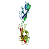
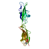
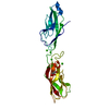
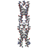
- Assembly
| Deposited unit | 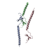
| ||||||||||||
|---|---|---|---|---|---|---|---|---|---|---|---|---|---|
| 1 | 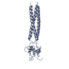
| ||||||||||||
| 2 | 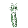
| ||||||||||||
| 3 | 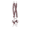
| ||||||||||||
| Unit cell |
| ||||||||||||
| Components on special symmetry positions |
|
-Components
| #1: Protein | Mass: 7981.825 Da / Num. of mol.: 3 Fragment: DELETION MUTANT M, RESIDUES 413 - 486 OF THE WILD TYPE Mutation: R416S, S421K, N425I, N428D, T433R Source method: isolated from a genetically manipulated source Source: (gene. exp.) Enterobacteria phage T4 (virus) / Genus: T4-like viruses / Species: Enterobacteria phage T4 sensu lato / Cell line: BL21 / Gene: WAC / Plasmid: BL21 / Species (production host): Escherichia coli / Production host:  #2: Water | ChemComp-HOH / |  Mass: 18.015 Da / Num. of mol.: 277 / Source method: isolated from a natural source / Formula: H2O Mass: 18.015 Da / Num. of mol.: 277 / Source method: isolated from a natural source / Formula: H2O |
|---|
-Experimental details
-Experiment
| Experiment | Method: X-RAY DIFFRACTION / Number of used crystals: 1 |
|---|
- Sample preparation
| Crystal | Density Matthews: 2.18 Å3/Da / Density % sol: 45 % | ||||||||||||||||||||||||||||||||||||||||||
|---|---|---|---|---|---|---|---|---|---|---|---|---|---|---|---|---|---|---|---|---|---|---|---|---|---|---|---|---|---|---|---|---|---|---|---|---|---|---|---|---|---|---|---|
| Crystal grow | Method: vapor diffusion, hanging drop / pH: 7.5 Details: HANGING DROPS WITH 20MG/ML PROTEIN AND 1.75M LI2SO4, 0.1M TRIS-HCL, PH7.5, AS PRECIPITANT, vapor diffusion - hanging drop | ||||||||||||||||||||||||||||||||||||||||||
| Crystal grow | *PLUS Method: vapor diffusion, hanging drop | ||||||||||||||||||||||||||||||||||||||||||
| Components of the solutions | *PLUS
|
-Data collection
| Diffraction | Mean temperature: 100 K |
|---|---|
| Diffraction source | Source: ROTATING ANODE / Type: RIGAKU / Wavelength: 1.5418 |
| Detector | Type: RIGAKU RAXIS IIC / Detector: IMAGE PLATE / Date: Sep 1, 1995 / Details: MIRRORS |
| Radiation | Monochromator: NI FILTER / Monochromatic (M) / Laue (L): M / Scattering type: x-ray |
| Radiation wavelength | Wavelength: 1.5418 Å / Relative weight: 1 |
| Reflection | Resolution: 1.85→23 Å / Num. obs: 14861 / % possible obs: 89.8 % / Observed criterion σ(I): 0 / Redundancy: 2.7 % / Biso Wilson estimate: 20.9 Å2 / Rsym value: 0.036 |
| Reflection shell | Resolution: 1.85→1.91 Å / Rsym value: 0.182 / % possible all: 53.6 |
| Reflection | *PLUS Num. measured all: 39783 / Rmerge(I) obs: 0.036 |
| Reflection shell | *PLUS % possible obs: 53.6 % / Rmerge(I) obs: 0.182 |
- Processing
| Software |
| ||||||||||||||||||||||||||||||||||||||||||||||||||||||||||||
|---|---|---|---|---|---|---|---|---|---|---|---|---|---|---|---|---|---|---|---|---|---|---|---|---|---|---|---|---|---|---|---|---|---|---|---|---|---|---|---|---|---|---|---|---|---|---|---|---|---|---|---|---|---|---|---|---|---|---|---|---|---|
| Refinement | Method to determine structure: MOLECULAR REPLACEMENT Starting model: PDB ENTRY 1AA0 Resolution: 1.85→23 Å / Isotropic thermal model: RESTRAINED / Cross valid method: THROUGHOUT / σ(F): 0 Details: BULK SOLVENT WAS MODELED USING A STANDARD X-PLOR PROCEDURE, WITH BULK SOLVENT DENSITY 0.432 E/A3.
| ||||||||||||||||||||||||||||||||||||||||||||||||||||||||||||
| Displacement parameters | Biso mean: 32.81 Å2 | ||||||||||||||||||||||||||||||||||||||||||||||||||||||||||||
| Refinement step | Cycle: LAST / Resolution: 1.85→23 Å
| ||||||||||||||||||||||||||||||||||||||||||||||||||||||||||||
| Refine LS restraints |
| ||||||||||||||||||||||||||||||||||||||||||||||||||||||||||||
| LS refinement shell | Resolution: 1.85→1.93 Å / Rfactor Rfree: 0.416 / Rfactor Rwork: 0.317 / Total num. of bins used: 10 | ||||||||||||||||||||||||||||||||||||||||||||||||||||||||||||
| Xplor file |
| ||||||||||||||||||||||||||||||||||||||||||||||||||||||||||||
| Software | *PLUS Name: X-PLOR / Version: 3.8 / Classification: refinement | ||||||||||||||||||||||||||||||||||||||||||||||||||||||||||||
| Refinement | *PLUS | ||||||||||||||||||||||||||||||||||||||||||||||||||||||||||||
| Solvent computation | *PLUS | ||||||||||||||||||||||||||||||||||||||||||||||||||||||||||||
| Displacement parameters | *PLUS | ||||||||||||||||||||||||||||||||||||||||||||||||||||||||||||
| Refine LS restraints | *PLUS
| ||||||||||||||||||||||||||||||||||||||||||||||||||||||||||||
| LS refinement shell | *PLUS Rfactor obs: 0.317 |
