 Movie
Movie Controller


+ Open data

- Basic information
| Entry | Database: PDB / ID: 3lnf | ||||||
|---|---|---|---|---|---|---|---|
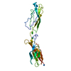
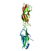
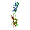
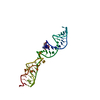
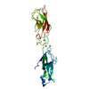
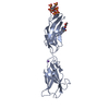
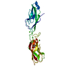
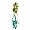
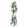
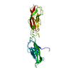
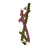
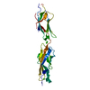
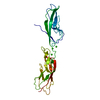
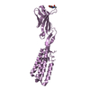
| Title | Crystal structure of E-cadherin EC12 K14EW2A | ||||||
 Components Components | Cadherin-1 | ||||||
 Keywords Keywords | CELL ADHESION / cadherin / Calcium / Cell junction / Cell membrane / Glycoprotein / Transmembrane | ||||||
| Function / homology |  Function and homology information Function and homology informationuterine epithelium development / Apoptotic cleavage of cell adhesion proteins / Regulation of CDH1 posttranslational processing and trafficking to plasma membrane / desmosome assembly / salivary gland cavitation / regulation of branching involved in salivary gland morphogenesis / Adherens junctions interactions / Degradation of the extracellular matrix / RHO GTPases activate IQGAPs / Integrin cell surface interactions ...uterine epithelium development / Apoptotic cleavage of cell adhesion proteins / Regulation of CDH1 posttranslational processing and trafficking to plasma membrane / desmosome assembly / salivary gland cavitation / regulation of branching involved in salivary gland morphogenesis / Adherens junctions interactions / Degradation of the extracellular matrix / RHO GTPases activate IQGAPs / Integrin cell surface interactions / Regulation of CDH1 Function / Degradation of CDH1 / gamma-catenin binding / desmosome / positive regulation of cell-cell adhesion / lateral loop / regulation of protein localization to cell surface / negative regulation of axon extension / cell-cell adhesion mediator activity / alpha-catenin binding / cellular response to indole-3-methanol / trophectodermal cell differentiation / calcium-dependent cell-cell adhesion / intestinal epithelial cell development / bicellular tight junction assembly / Schmidt-Lanterman incisure / regulation of protein catabolic process at postsynapse, modulating synaptic transmission / flotillin complex / cell-cell adhesion mediated by cadherin / adherens junction organization / epithelial cell morphogenesis / catenin complex / regulation of neuron migration / node of Ranvier / negative regulation of protein processing / cell-cell junction assembly / GTPase activating protein binding / negative regulation of cell-cell adhesion / ankyrin binding / cellular response to lithium ion / apical junction complex / homophilic cell-cell adhesion / decidualization / microvillus / establishment of skin barrier / cochlea development / lateral plasma membrane / negative regulation of protein localization to plasma membrane / positive regulation of protein localization / cytoskeletal protein binding / synapse assembly / embryo implantation / cell adhesion molecule binding / protein tyrosine kinase binding / axon terminus / negative regulation of cell migration / cell periphery / protein localization to plasma membrane / adherens junction / trans-Golgi network / cellular response to amino acid stimulus / negative regulation of canonical Wnt signaling pathway / sensory perception of sound / cell-cell adhesion / positive regulation of protein import into nucleus / beta-catenin binding / cell morphogenesis / negative regulation of epithelial cell proliferation / cytoplasmic side of plasma membrane / apical part of cell / cell-cell junction / cell junction / cell migration / regulation of protein localization / lamellipodium / actin cytoskeleton / regulation of gene expression / actin cytoskeleton organization / protein phosphatase binding / molecular adaptor activity / in utero embryonic development / basolateral plasma membrane / endosome / postsynapse / cadherin binding / protein domain specific binding / axon / calcium ion binding / positive regulation of DNA-templated transcription / perinuclear region of cytoplasm / glutamatergic synapse / cell surface / membrane / identical protein binding / plasma membrane / cytoplasm Similarity search - Function | ||||||
| Biological species |  | ||||||
| Method |  X-RAY DIFFRACTION / SYNCHROTRON / MOLECULAR REPLACEMENT / Resolution: 2.5 Å X-RAY DIFFRACTION / SYNCHROTRON / MOLECULAR REPLACEMENT / Resolution: 2.5 Å | ||||||
 Authors Authors | Jin, X. / Harrison, O. / Shapiro, L. | ||||||
 Citation Citation | Journal: Nat.Struct.Mol.Biol. / Year: 2010 Title: Two-step adhesive binding by classical cadherins. Authors: Harrison, O.J. / Bahna, F. / Katsamba, P.S. / Jin, X. / Brasch, J. / Vendome, J. / Ahlsen, G. / Carroll, K.J. / Price, S.R. / Honig, B. / Shapiro, L. | ||||||
| History |
|
- Structure visualization
| Structure viewer | Molecule: MolmilJmol/JSmol |
|---|
- Downloads & links
-Download
| PDBx/mmCIF format | 3lnf.cif.gz | 96 KB | Display | PDBx/mmCIF format |
|---|---|---|---|---|
| PDB format | pdb3lnf.ent.gz | 72.5 KB | Display | PDB format |
| PDBx/mmJSON format | 3lnf.json.gz | Tree view | PDBx/mmJSON format | |
| Others |  Other downloads Other downloads |
-Validation report
| Arichive directory | https://data.pdbj.org/pub/pdb/validation_reports/ln/3lnfftp://data.pdbj.org/pub/pdb/validation_reports/ln/3lnf | HTTPS FTP |
|---|
-Related structure data
| Related structure data |  3lndC  3lneC  3lngC  3lnhC  3lniC 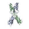 1edhS C: citing same article ( S: Starting model for refinement |
|---|---|
| Similar structure data |


-Links
 PDBj
PDBj
- Assembly
| Deposited unit | 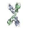
| ||||||||
|---|---|---|---|---|---|---|---|---|---|
| 1 | 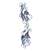
| ||||||||
| 2 | 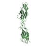
| ||||||||
| Unit cell |
|
-Components
| #1: Protein | Mass: 23145.668 Da / Num. of mol.: 2 / Fragment: UNP residues 157-369 / Mutation: K14E,W2A Source method: isolated from a genetically manipulated source Source: (gene. exp.)  #2: Chemical | ChemComp-CA /   Mass: 40.078 Da / Num. of mol.: 6 / Source method: obtained synthetically / Formula: Ca Mass: 40.078 Da / Num. of mol.: 6 / Source method: obtained synthetically / Formula: Ca#3: Water | ChemComp-HOH / |  Mass: 18.015 Da / Num. of mol.: 169 / Source method: isolated from a natural source / Formula: H2O Mass: 18.015 Da / Num. of mol.: 169 / Source method: isolated from a natural source / Formula: H2O |
|---|
-Experimental details
-Experiment
| Experiment | Method: X-RAY DIFFRACTION / Number of used crystals: 1 |
|---|
- Sample preparation
| Crystal | Density Matthews: 2.94 Å3/Da / Density % sol: 58.19 % |
|---|---|
| Crystal grow | Temperature: 293 K / Method: vapor diffusion, hanging drop / pH: 8.5 Details: 1.2M ammonium sulfate, 0.1M Tris-Cl pH 8.5, 15% (v/v) glycerol, VAPOR DIFFUSION, HANGING DROP, temperature 293K |
-Data collection
| Diffraction | Mean temperature: 100 K |
|---|---|
| Diffraction source | Source: SYNCHROTRON / Site: NSLS  / Beamline: X4A / Wavelength: 0.97897 Å / Beamline: X4A / Wavelength: 0.97897 Å |
| Detector | Type: ADSC QUANTUM 4 / Detector: CCD / Date: Feb 16, 2008 |
| Radiation | Monochromator: Si 111 CHANNEL / Protocol: SINGLE WAVELENGTH / Monochromatic (M) / Laue (L): M / Scattering type: x-ray |
| Radiation wavelength | Wavelength: 0.97897 Å / Relative weight: 1 |
| Reflection | Resolution: 2.5→50 Å / Num. all: 17934 / Num. obs: 17683 / % possible obs: 98.6 % / Observed criterion σ(F): 1 / Observed criterion σ(I): 1 / Redundancy: 2.5 % / Rmerge(I) obs: 0.111 / Net I/σ(I): 8.2 |
- Processing
| Software |
| |||||||||||||||||||||||||||||||||||||||||||||||||
|---|---|---|---|---|---|---|---|---|---|---|---|---|---|---|---|---|---|---|---|---|---|---|---|---|---|---|---|---|---|---|---|---|---|---|---|---|---|---|---|---|---|---|---|---|---|---|---|---|---|---|
| Refinement | Method to determine structure: MOLECULAR REPLACEMENT Starting model: PDB ENTRY 1EDH Resolution: 2.5→28.635 Å / SU ML: 0.36 / σ(F): 1.97 / Stereochemistry target values: ML
| |||||||||||||||||||||||||||||||||||||||||||||||||
| Solvent computation | Shrinkage radii: 0.9 Å / VDW probe radii: 1.11 Å / Solvent model: FLAT BULK SOLVENT MODEL / Bsol: 21.401 Å2 / ksol: 0.369 e/Å3 | |||||||||||||||||||||||||||||||||||||||||||||||||
| Refinement step | Cycle: LAST / Resolution: 2.5→28.635 Å
| |||||||||||||||||||||||||||||||||||||||||||||||||
| Refine LS restraints |
| |||||||||||||||||||||||||||||||||||||||||||||||||
| LS refinement shell |
|
