 Movie
Movie Controller


+ Open data

- Basic information
| Entry | Database: PDB / ID: 5ckq | |||||||||
|---|---|---|---|---|---|---|---|---|---|---|
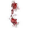
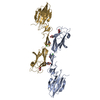
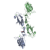
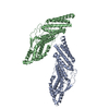
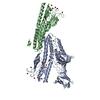
| Title | CUB1-EGF-CUB2 domains of rat MASP-1 | |||||||||
 Components Components | Mannan-binding lectin serine protease 1 | |||||||||
 Keywords Keywords | HYDROLASE / CUB1-EGF-CUB2 / serine protease / lectin pathway / complement | |||||||||
| Function / homology |  Function and homology information Function and homology informationFicolins bind to repetitive carbohydrate structures on the target cell surface / Lectin pathway of complement activation / Initial triggering of complement / activation of membrane attack complex / negative regulation of complement activation / complement activation, lectin pathway / opsonization / symbiont cell surface / complement activation, alternative pathway / complement activation ...Ficolins bind to repetitive carbohydrate structures on the target cell surface / Lectin pathway of complement activation / Initial triggering of complement / activation of membrane attack complex / negative regulation of complement activation / complement activation, lectin pathway / opsonization / symbiont cell surface / complement activation, alternative pathway / complement activation / zymogen activation / Hydrolases; Acting on peptide bonds (peptidases); Serine endopeptidases / protein maturation / calcium-dependent protein binding / peptidase activity / killing of cells of another organism / serine-type endopeptidase activity / calcium ion binding / protein homodimerization activity / : / nucleoplasm / identical protein binding / cytosol Similarity search - Function | |||||||||
| Biological species |  | |||||||||
| Method |  X-RAY DIFFRACTION / SYNCHROTRON / MOLECULAR REPLACEMENT / molecular replacement / Resolution: 3.704 Å X-RAY DIFFRACTION / SYNCHROTRON / MOLECULAR REPLACEMENT / molecular replacement / Resolution: 3.704 Å | |||||||||
 Authors Authors | Nan, R. / Furze, C.M. / Wright, D.W. / Gor, J. / Wallis, R. / Perkins, S.J. | |||||||||
| Funding support |  United Kingdom, 2items United Kingdom, 2items
| |||||||||
 Citation Citation | Journal: Structure / Year: 2017 Title: Flexibility in Mannan-Binding Lectin-Associated Serine Proteases-1 and -2 Provides Insight on Lectin Pathway Activation. Authors: Nan, R. / Furze, C.M. / Wright, D.W. / Gor, J. / Wallis, R. / Perkins, S.J. | |||||||||
| History |
|
- Structure visualization
| Structure viewer | Molecule: MolmilJmol/JSmol |
|---|
- Downloads & links
-Download
| PDBx/mmCIF format | 5ckq.cif.gz | 131.5 KB | Display | PDBx/mmCIF format |
|---|---|---|---|---|
| PDB format | pdb5ckq.ent.gz | 103.6 KB | Display | PDB format |
| PDBx/mmJSON format | 5ckq.json.gz | Tree view | PDBx/mmJSON format | |
| Others |  Other downloads Other downloads |
-Validation report
| Arichive directory | https://data.pdbj.org/pub/pdb/validation_reports/ck/5ckqftp://data.pdbj.org/pub/pdb/validation_reports/ck/5ckq | HTTPS FTP |
|---|
-Related structure data
| Related structure data | 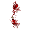 5cisC  5ckmC 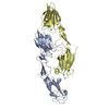 5cknC  3demS S: Starting model for refinement C: citing same article ( |
|---|---|
| Similar structure data |

-Links
 PDBj
PDBj


- Assembly
| Deposited unit | 
| ||||||||
|---|---|---|---|---|---|---|---|---|---|
| 1 | 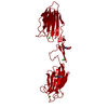
| ||||||||
| Unit cell |
|
-Components
| #1: Protein | Mass: 31889.996 Da / Num. of mol.: 1 Source method: isolated from a genetically manipulated source Source: (gene. exp.)  Cricetulus griseus (Chinese hamster) Cricetulus griseus (Chinese hamster)References: UniProt: Q8CHN8, Hydrolases; Acting on peptide bonds (peptidases); Serine endopeptidases | ||||||
|---|---|---|---|---|---|---|---|
| #2: Sugar |   Type: D-saccharide, beta linking / Mass: 221.208 Da / Num. of mol.: 2 Type: D-saccharide, beta linking / Mass: 221.208 Da / Num. of mol.: 2Source method: isolated from a genetically manipulated source Formula: C8H15NO6 #3: Chemical |   Mass: 40.078 Da / Num. of mol.: 3 / Source method: obtained synthetically / Formula: Ca Mass: 40.078 Da / Num. of mol.: 3 / Source method: obtained synthetically / Formula: Ca#4: Chemical | ChemComp-NA / |   Mass: 22.990 Da / Num. of mol.: 1 / Source method: obtained synthetically / Formula: Na Mass: 22.990 Da / Num. of mol.: 1 / Source method: obtained synthetically / Formula: NaHas protein modification | Y | |
-Experimental details
-Experiment
| Experiment | Method: X-RAY DIFFRACTION / Number of used crystals: 1 |
|---|
- Sample preparation
| Crystal | Density Matthews: 4.65 Å3/Da / Density % sol: 73.57 % |
|---|---|
| Crystal grow | Temperature: 293 K / Method: vapor diffusion, sitting drop / pH: 6.5 Details: 100 mM imidazole/MOPS pH 6.5 containing 20% ethylene glycol and 10% PEG8K |
-Data collection
| Diffraction | Mean temperature: 100 K |
|---|---|
| Diffraction source | Source: SYNCHROTRON / Site: Diamond / Beamline: I04 / Wavelength: 0.9795 Å |
| Detector | Type: DECTRIS PILATUS 2M / Detector: PIXEL / Date: May 13, 2013 |
| Radiation | Protocol: SINGLE WAVELENGTH / Monochromatic (M) / Laue (L): M / Scattering type: x-ray |
| Radiation wavelength | Wavelength: 0.9795 Å / Relative weight: 1 |
| Reflection | Resolution: 3.7→76.4 Å / Num. obs: 6473 / % possible obs: 99.9 % / Redundancy: 5.6 % / Rsym value: 0.082 / Net I/σ(I): 9.2 |
| Reflection shell | Resolution: 3.7→4.14 Å / Redundancy: 5.5 % / Rmerge(I) obs: 0.742 / Mean I/σ(I) obs: 2.1 / % possible all: 99.9 |
-Phasing
| Phasing | Method: molecular replacement |
|---|
- Processing
| Software |
| ||||||||||||||||||||||||||||||||||||||||||||||||||||||||||||||||||||||||||||||||||||||||||||||||||||
|---|---|---|---|---|---|---|---|---|---|---|---|---|---|---|---|---|---|---|---|---|---|---|---|---|---|---|---|---|---|---|---|---|---|---|---|---|---|---|---|---|---|---|---|---|---|---|---|---|---|---|---|---|---|---|---|---|---|---|---|---|---|---|---|---|---|---|---|---|---|---|---|---|---|---|---|---|---|---|---|---|---|---|---|---|---|---|---|---|---|---|---|---|---|---|---|---|---|---|---|---|---|
| Refinement | Method to determine structure: MOLECULAR REPLACEMENT Starting model: 3DEM Resolution: 3.704→76.355 Å / SU ML: 0.38 / Cross valid method: FREE R-VALUE / σ(F): 1.36 / Phase error: 39.03 / Stereochemistry target values: ML
| ||||||||||||||||||||||||||||||||||||||||||||||||||||||||||||||||||||||||||||||||||||||||||||||||||||
| Solvent computation | Shrinkage radii: 0.9 Å / VDW probe radii: 1.11 Å / Solvent model: FLAT BULK SOLVENT MODEL | ||||||||||||||||||||||||||||||||||||||||||||||||||||||||||||||||||||||||||||||||||||||||||||||||||||
| Displacement parameters | Biso max: 453.92 Å2 / Biso mean: 187.1629 Å2 / Biso min: 108.93 Å2 | ||||||||||||||||||||||||||||||||||||||||||||||||||||||||||||||||||||||||||||||||||||||||||||||||||||
| Refinement step | Cycle: final / Resolution: 3.704→76.355 Å
| ||||||||||||||||||||||||||||||||||||||||||||||||||||||||||||||||||||||||||||||||||||||||||||||||||||
| Refine LS restraints |
| ||||||||||||||||||||||||||||||||||||||||||||||||||||||||||||||||||||||||||||||||||||||||||||||||||||
| LS refinement shell | Refine-ID: X-RAY DIFFRACTION / Total num. of bins used: 2 / % reflection obs: 100 %
| ||||||||||||||||||||||||||||||||||||||||||||||||||||||||||||||||||||||||||||||||||||||||||||||||||||
| Refinement TLS params. | Method: refined / Refine-ID: X-RAY DIFFRACTION
| ||||||||||||||||||||||||||||||||||||||||||||||||||||||||||||||||||||||||||||||||||||||||||||||||||||
| Refinement TLS group |
|
