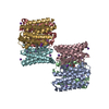
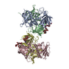
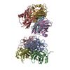
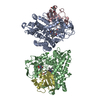
[English] 日本語
Yorodumi
- PDB-6elc: Crystal Structure of O-linked Glycosylated VSG3 -

+
Open data


ID or keywords:

Loading...

-
Basic information

Entry
Database: PDB / ID: 6elc
TitleCrystal Structure of O-linked Glycosylated VSG3
ComponentsVariant surface glycoprotein
KeywordsIMMUNE SYSTEM / trypanosome / VSG / sleeping sickness / antigen
Function / homologyTrypanosome variant surface glycoprotein, B-type, N-terminal domain / Trypanosomal VSG domain / Trypanosome variant surface glycoprotein, C-terminal / Trypanosome variant surface glycoprotein C-terminal domain / side of membrane / plasma membrane / alpha-D-glucopyranose / Variant surface glycoprotein
Function and homology information
Biological speciesTrypanosoma brucei brucei (eukaryote)
MethodX-RAY DIFFRACTION / SYNCHROTRON / MOLECULAR REPLACEMENT / Resolution: 1.41 Å
AuthorsStebbins, C.E.
Funding support United States, United Kingdom, 5items
OrganizationGrant numberCountry
National Institutes of Health/National Institute of General Medical Sciences (NIH/NIGMS)GM103403 United States
National Institutes of Health/National Institute Of Allergy and Infectious Diseases (NIH/NIAID)AI085973 United States
Wellcome Trust101842 United Kingdom
Wellcome Trust097045 United Kingdom
National Institutes of Health United States
CitationJournal: Nat Microbiol / Year: 2018
Title: African trypanosomes evade immune clearance by O-glycosylation of the VSG surface coat.
Authors: Pinger, J. / Nesic, D. / Ali, L. / Aresta-Branco, F. / Lilic, M. / Chowdhury, S. / Kim, H.S. / Verdi, J. / Raper, J. / Ferguson, M.A.J. / Papavasiliou, F.N. / Stebbins, C.E.
History
DepositionSep 28, 2017Deposition site: PDBE / Processing site: PDBE
Revision 1.0Jul 11, 2018Provider: repository / Type: Initial release
Revision 1.1Jul 18, 2018Group: Data collection / Database references / Category: citation / citation_author / Item: _citation.pdbx_database_id_PubMed / _citation.title
Revision 1.2Aug 29, 2018Group: Data collection / Database references / Category: citation / citation_author
Item: _citation.journal_volume / _citation.page_first ..._citation.journal_volume / _citation.page_first / _citation.page_last / _citation_author.identifier_ORCID
Revision 2.0Jul 29, 2020Group: Atomic model / Data collection ...Atomic model / Data collection / Derived calculations / Structure summary
Category: atom_site / atom_site_anisotrop ...atom_site / atom_site_anisotrop / chem_comp / entity / pdbx_branch_scheme / pdbx_chem_comp_identifier / pdbx_entity_branch / pdbx_entity_branch_descriptor / pdbx_entity_branch_link / pdbx_entity_branch_list / pdbx_entity_nonpoly / pdbx_nonpoly_scheme / pdbx_struct_assembly_gen / pdbx_struct_special_symmetry / struct_asym / struct_conn / struct_site / struct_site_gen
Item: _atom_site.auth_asym_id / _atom_site.auth_seq_id ..._atom_site.auth_asym_id / _atom_site.auth_seq_id / _atom_site.label_asym_id / _atom_site.label_entity_id / _atom_site_anisotrop.pdbx_auth_asym_id / _atom_site_anisotrop.pdbx_auth_seq_id / _atom_site_anisotrop.pdbx_label_asym_id / _chem_comp.name / _chem_comp.type / _pdbx_struct_assembly_gen.asym_id_list / _pdbx_struct_special_symmetry.label_asym_id / _struct_conn.pdbx_role / _struct_conn.ptnr1_auth_asym_id / _struct_conn.ptnr1_auth_seq_id / _struct_conn.ptnr1_label_asym_id / _struct_conn.ptnr2_auth_asym_id / _struct_conn.ptnr2_auth_seq_id / _struct_conn.ptnr2_label_asym_id
Description: Carbohydrate remediation / Provider: repository / Type: Remediation
Revision 2.1Mar 30, 2022Group: Author supporting evidence / Database references ...Author supporting evidence / Database references / Derived calculations / Structure summary
Category: chem_comp / database_2 ...chem_comp / database_2 / pdbx_audit_support / struct_conn
Item: _chem_comp.pdbx_synonyms / _database_2.pdbx_DOI ..._chem_comp.pdbx_synonyms / _database_2.pdbx_DOI / _database_2.pdbx_database_accession / _pdbx_audit_support.funding_organization / _struct_conn.pdbx_leaving_atom_flag
Revision 2.2Oct 16, 2024Group: Data collection / Structure summary
Category: chem_comp_atom / chem_comp_bond ...chem_comp_atom / chem_comp_bond / pdbx_entry_details / pdbx_modification_feature

-
Structure visualization

Structure viewerMolecule:
MolmilJmol/JSmol

Downloads & links

-
Assembly

Deposited unit
A: Variant surface glycoprotein
hetero molecules


Theoretical massNumber of molelcules
Total (without water)54,8633
Polymers53,9341
Non-polymers9292
Water8,557475
1
A: Variant surface glycoprotein
hetero molecules

A: Variant surface glycoprotein
hetero molecules

A: Variant surface glycoprotein
hetero molecules


Theoretical massNumber of molelcules
Total (without water)164,5909
Polymers161,8033
Non-polymers2,7876
Water543
TypeNameSymmetry operationNumber
identity operation1_555x,y,z1
crystal symmetry operation5_555z,x,y1
crystal symmetry operation9_555y,z,x1
Buried area14420 Å2
ΔGint12 kcal/mol
Surface area41540 Å2
MethodPISA
Unit cell
Length a, b, c (Å)129.854, 129.854, 129.854
Angle α, β, γ (deg.)90.00, 90.00, 90.00
Int Tables number199
Space group name H-MI213
Components on special symmetry positions
IDModelComponents
11A-1053-

HOH

21A-1116-

HOH

31A-1137-

HOH

41A-1145-

HOH

51A-1162-

HOH

61A-1170-

HOH

71A-1171-

HOH

81A-1175-

HOH

-
Components

#1: Protein Variant surface glycoprotein / VSG3 MITat1.3 VSG224


Mass: 53934.395 Da / Num. of mol.: 1 / Source method: isolated from a natural source / Source: (natural) Trypanosoma brucei brucei (eukaryote) / References: UniProt: B3GVK1
#2: Polysaccharide alpha-D-mannopyranose-(1-6)-beta-D-mannopyranose-(1-4)-2-acetamido-2-deoxy-beta-D-glucopyranose-(1- ...alpha-D-mannopyranose-(1-6)-beta-D-mannopyranose-(1-4)-2-acetamido-2-deoxy-beta-D-glucopyranose-(1-4)-2-acetamido-2-deoxy-beta-D-glucopyranose


Type: oligosaccharide / Mass: 748.682 Da / Num. of mol.: 1
Source method: isolated from a genetically manipulated source
DescriptorTypeProgram
DManpa1-6DManpb1-4DGlcpNAcb1-4DGlcpNAcb1-Glycam Condensed SequenceGMML 1.0
WURCS=2.0/3,4,3/[a2122h-1b_1-5_2*NCC/3=O][a1122h-1b_1-5][a1122h-1a_1-5]/1-1-2-3/a4-b1_b4-c1_c6-d1WURCSPDB2Glycan 1.1.0
[]{[(4+1)][b-D-GlcpNAc]{[(4+1)][b-D-GlcpNAc]{[(4+1)][b-D-Manp]{[(6+1)][a-D-Manp]{}}}}}LINUCSPDB-CARE
#3: Sugar ChemComp-GLC / alpha-D-glucopyranose / alpha-D-glucose / D-glucose / glucose


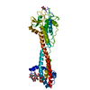
Type: D-saccharide, alpha linking / Mass: 180.156 Da / Num. of mol.: 1
Source method: isolated from a genetically manipulated source
Formula: C6H12O6
IdentifierTypeProgram
DGlcpaCONDENSED IUPAC CARBOHYDRATE SYMBOLGMML 1.0
a-D-glucopyranoseCOMMON NAMEGMML 1.0
a-D-GlcpIUPAC CARBOHYDRATE SYMBOLPDB-CARE 1.0
GlcSNFG CARBOHYDRATE SYMBOLGMML 1.0
#4: Water ChemComp-HOH / water


Mass: 18.015 Da / Num. of mol.: 475 / Source method: isolated from a natural source / Formula: H2O
Has protein modificationY

-
Experimental details

-
Experiment

ExperimentMethod: X-RAY DIFFRACTION / Number of used crystals: 1

-
Sample preparation

Crystal growTemperature: 298 K / Method: vapor diffusion, hanging drop / pH: 7.5
Details: Hanging drops formed from mixing a 1:1 volume ratio of the protein with an equilibration buffer consisting of 16-22% polyethylene glycol (PEG) molecular weight 3350 Da, 200-325mM NaCl, and ...Details: Hanging drops formed from mixing a 1:1 volume ratio of the protein with an equilibration buffer consisting of 16-22% polyethylene glycol (PEG) molecular weight 3350 Da, 200-325mM NaCl, and 100mM Tris pH 8.2 (tris(hydroxymethyl)aminomethane)

-
Data collection

DiffractionMean temperature: 100 K
Diffraction sourceSource: SYNCHROTRON / Site: APS / Beamline: 24-ID-C / Wavelength: 0.9194 Å
DetectorType: DECTRIS PILATUS 6M-F / Detector: PIXEL / Date: Aug 10, 2015
RadiationProtocol: SINGLE WAVELENGTH / Monochromatic (M) / Laue (L): M / Scattering type: x-ray
Radiation wavelengthWavelength: 0.9194 Å / Relative weight: 1
ReflectionResolution: 1.41→91.82 Å / Num. obs: 68683 / % possible obs: 97.9 % / Observed criterion σ(I): 19.1 / Redundancy: 4.1 % / CC1/2: 0.999 / Rpim(I) all: 0.022 / Net I/σ(I): 19.1
Reflection shellResolution: 1.41→1.43 Å / Redundancy: 1.6 % / Mean I/σ(I) obs: 0.6 / Num. unique obs: 2732 / CC1/2: 0.407 / Rpim(I) all: 0.725 / % possible all: 73.9

-
Processing

Software
NameVersionClassification
REFMAC5.8.0135refinement
XDSJune 17, 2015data reduction
XDSJune 17, 2015data scaling
RefinementMethod to determine structure: MOLECULAR REPLACEMENT / Resolution: 1.41→91.82 Å / Cor.coef. Fo:Fc: 0.979 / Cor.coef. Fo:Fc free: 0.968 / SU B: 2.036 / SU ML: 0.036 / Cross valid method: THROUGHOUT / ESU R: 0.056 / ESU R Free: 0.054 / Details: HYDROGENS HAVE BEEN ADDED IN THE RIDING POSITIONS
RfactorNum. reflection% reflectionSelection details
Rfree0.16921 3462 5.1 %RANDOM
Rwork0.13003 ---
obs0.13193 64954 97.9 %-
Solvent computationIon probe radii: 0.8 Å / Shrinkage radii: 0.8 Å / VDW probe radii: 1.2 Å
Displacement parametersBiso mean: 23.203 Å2
Baniso -1Baniso -2Baniso -3
1-0 Å20 Å20 Å2
2--0 Å20 Å2
3---0 Å2
Refinement stepCycle: 1 / Resolution: 1.41→91.82 Å
ProteinNucleic acidLigandSolventTotal
Num. atoms2652 0 61 475 3188
Refine LS restraints
Refine-IDTypeDev idealDev ideal targetNumber
X-RAY DIFFRACTIONr_bond_refined_d0.0170.022762
X-RAY DIFFRACTIONr_bond_other_d0.0020.022613
X-RAY DIFFRACTIONr_angle_refined_deg1.7581.9773761
X-RAY DIFFRACTIONr_angle_other_deg1.05136026
X-RAY DIFFRACTIONr_dihedral_angle_1_deg5.2475354
X-RAY DIFFRACTIONr_dihedral_angle_2_deg34.75325.877114
X-RAY DIFFRACTIONr_dihedral_angle_3_deg12.62715445
X-RAY DIFFRACTIONr_dihedral_angle_4_deg19.2681512
X-RAY DIFFRACTIONr_chiral_restr0.1210.2448
X-RAY DIFFRACTIONr_gen_planes_refined0.010.023148
X-RAY DIFFRACTIONr_gen_planes_other0.0020.02586
X-RAY DIFFRACTIONr_nbd_refined
X-RAY DIFFRACTIONr_nbd_other
X-RAY DIFFRACTIONr_nbtor_refined
X-RAY DIFFRACTIONr_nbtor_other
X-RAY DIFFRACTIONr_xyhbond_nbd_refined
X-RAY DIFFRACTIONr_xyhbond_nbd_other
X-RAY DIFFRACTIONr_metal_ion_refined
X-RAY DIFFRACTIONr_metal_ion_other
X-RAY DIFFRACTIONr_symmetry_vdw_refined
X-RAY DIFFRACTIONr_symmetry_vdw_other
X-RAY DIFFRACTIONr_symmetry_hbond_refined
X-RAY DIFFRACTIONr_symmetry_hbond_other
X-RAY DIFFRACTIONr_symmetry_metal_ion_refined
X-RAY DIFFRACTIONr_symmetry_metal_ion_other
X-RAY DIFFRACTIONr_mcbond_it2.5531.9311422
X-RAY DIFFRACTIONr_mcbond_other2.5531.9291421
X-RAY DIFFRACTIONr_mcangle_it3.1572.9051774
X-RAY DIFFRACTIONr_mcangle_other3.1562.9061775
X-RAY DIFFRACTIONr_scbond_it3.7172.4071340
X-RAY DIFFRACTIONr_scbond_other3.7172.4071340
X-RAY DIFFRACTIONr_scangle_it
X-RAY DIFFRACTIONr_scangle_other4.3323.4571988
X-RAY DIFFRACTIONr_long_range_B_refined4.60418.6963706
X-RAY DIFFRACTIONr_long_range_B_other4.2817.6893454
X-RAY DIFFRACTIONr_rigid_bond_restr2.91735373
X-RAY DIFFRACTIONr_sphericity_free30.2475117
X-RAY DIFFRACTIONr_sphericity_bonded11.40255684
LS refinement shellResolution: 1.41→1.446 Å / Total num. of bins used: 20
RfactorNum. reflection% reflection
Rfree0.263 215 -
Rwork0.24 3859 -
obs--79.38 %

+
About Yorodumi

-
News

-
Feb 9, 2022. New format data for meta-information of EMDB entries

New format data for meta-information of EMDB entries

  • Version 3 of the EMDB header file is now the official format.
  • The previous official version 1.9 will be removed from the archive.

Related info.:EMDB header

External links:wwPDB to switch to version 3 of the EMDB data model

-
Aug 12, 2020. Covid-19 info

Covid-19 info

URL: https://pdbj.org/emnavi/covid19.php

New page: Covid-19 featured information page in EM Navigator.

Related info.:Covid-19 info / Mar 5, 2020. Novel coronavirus structure data

+
Mar 5, 2020. Novel coronavirus structure data

Novel coronavirus structure data

Related info.:Yorodumi Speices / Aug 12, 2020. Covid-19 info

External links:COVID-19 featured content - PDBj / Molecule of the Month (242):Coronavirus Proteases

+
Jan 31, 2019. EMDB accession codes are about to change! (news from PDBe EMDB page)

EMDB accession codes are about to change! (news from PDBe EMDB page)

  • The allocation of 4 digits for EMDB accession codes will soon come to an end. Whilst these codes will remain in use, new EMDB accession codes will include an additional digit and will expand incrementally as the available range of codes is exhausted. The current 4-digit format prefixed with “EMD-” (i.e. EMD-XXXX) will advance to a 5-digit format (i.e. EMD-XXXXX), and so on. It is currently estimated that the 4-digit codes will be depleted around Spring 2019, at which point the 5-digit format will come into force.
  • The EM Navigator/Yorodumi systems omit the EMD- prefix.

Related info.:Q: What is EMD? / ID/Accession-code notation in Yorodumi/EM Navigator

External links:EMDB Accession Codes are Changing Soon! / Contact to PDBj

+
Jul 12, 2017. Major update of PDB

Major update of PDB

  • wwPDB released updated PDB data conforming to the new PDBx/mmCIF dictionary.
  • This is a major update changing the version number from 4 to 5, and with Remediation, in which all the entries are updated.
  • In this update, many items about electron microscopy experimental information are reorganized (e.g. em_software).
  • Now, EM Navigator and Yorodumi are based on the updated data.

External links:wwPDB Remediation / Enriched Model Files Conforming to OneDep Data Standards Now Available in the PDB FTP Archive

-
Yorodumi

Thousand views of thousand structures

  • Yorodumi is a browser for structure data from EMDB, PDB, SASBDB, etc.
  • This page is also the successor to EM Navigator detail page, and also detail information page/front-end page for Omokage search.
  • The word "yorodu" (or yorozu) is an old Japanese word meaning "ten thousand". "mi" (miru) is to see.

Related info.:EMDB / PDB / SASBDB / Comparison of 3 databanks / Yorodumi Search / Aug 31, 2016. New EM Navigator & Yorodumi / Yorodumi Papers / Jmol/JSmol / Function and homology information / Changes in new EM Navigator and Yorodumi

Read more