 Movie
Movie Controller

[English] 日本語
 Yorodumi
- PDB-5fp2: Crystal structure of the siderophore receptor PirA from Pseudomon... -
+ Open data

- Basic information
| Entry | Database: PDB / ID: 5fp2 | ||||||
|---|---|---|---|---|---|---|---|
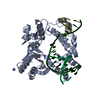
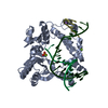
| Title | Crystal structure of the siderophore receptor PirA from Pseudomonas aeruginosa | ||||||
 Components Components | (FERRIC ENTEROBACTIN RECEPTOR PIRA) x 2 | ||||||
 Keywords Keywords | METAL TRANSPORT / TONB-DEPENDENT RECEPTOR / SIDEROPHORE RECEPTOR / OUTER-MEMBRANE PROTEIN | ||||||
| Function / homology |  Function and homology information Function and homology informationsiderophore transmembrane transport / siderophore uptake transmembrane transporter activity / siderophore transport / enterobactin transport / enterobactin transmembrane transporter activity / porin activity / pore complex / cell outer membrane / iron ion transport / signaling receptor activity Similarity search - Function | ||||||
| Biological species |   PSEUDOMONAS AERUGINOSA (bacteria) PSEUDOMONAS AERUGINOSA (bacteria) | ||||||
| Method |  X-RAY DIFFRACTION / SYNCHROTRON / MOLECULAR REPLACEMENT / Resolution: 2.97 Å X-RAY DIFFRACTION / SYNCHROTRON / MOLECULAR REPLACEMENT / Resolution: 2.97 Å | ||||||
 Authors Authors | Moynie, L. / Tortajada, A. / Naismith, J.H. | ||||||
 Citation Citation | Journal: Antimicrob. Agents Chemother. / Year: 2017 Title: Structure and Function of the PiuA and PirA Siderophore-Drug Receptors from Pseudomonas aeruginosa and Acinetobacter baumannii. Authors: Moynie, L. / Luscher, A. / Rolo, D. / Pletzer, D. / Tortajada, A. / Weingart, H. / Braun, Y. / Page, M.G. / Naismith, J.H. / Kohler, T. | ||||||
| History |
| ||||||
| Remark 700 | SHEET DETERMINATION METHOD: DSSP THE SHEETS PRESENTED AS "AC" IN EACH CHAIN ON SHEET RECORDS BELOW ... SHEET DETERMINATION METHOD: DSSP THE SHEETS PRESENTED AS "AC" IN EACH CHAIN ON SHEET RECORDS BELOW IS ACTUALLY AN 22-STRANDED BARREL THIS IS REPRESENTED BY A 23-STRANDED SHEET IN WHICH THE FIRST AND LAST STRANDS ARE IDENTICAL. SHEET DETERMINATION METHOD: DSSP THE SHEETS PRESENTED AS "BC" IN EACH CHAIN ON SHEET RECORDS BELOW IS ACTUALLY AN 22-STRANDED BARREL THIS IS REPRESENTED BY A 23-STRANDED SHEET IN WHICH THE FIRST AND LAST STRANDS ARE IDENTICAL. |
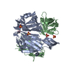
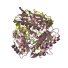
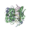
- Structure visualization
| Structure viewer | Molecule: MolmilJmol/JSmol |
|---|
- Downloads & links
-Download
| PDBx/mmCIF format | 5fp2.cif.gz | 423 KB | Display | PDBx/mmCIF format |
|---|---|---|---|---|
| PDB format | pdb5fp2.ent.gz | 352.7 KB | Display | PDB format |
| PDBx/mmJSON format | 5fp2.json.gz | Tree view | PDBx/mmJSON format | |
| Others |  Other downloads Other downloads |
-Validation report
| Arichive directory | https://data.pdbj.org/pub/pdb/validation_reports/fp/5fp2ftp://data.pdbj.org/pub/pdb/validation_reports/fp/5fp2 | HTTPS FTP |
|---|
-Related structure data
| Related structure data |  5fokC  5fp1C 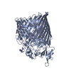 5fr8C  1fepS C: citing same article ( S: Starting model for refinement |
|---|---|
| Similar structure data |


-Links
 PDBj
PDBj

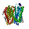
- Assembly
| Deposited unit | 
| ||||||||
|---|---|---|---|---|---|---|---|---|---|
| 1 | 
| ||||||||
| 2 | 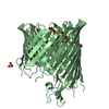
| ||||||||
| Unit cell |
| ||||||||
| Noncrystallographic symmetry (NCS) | NCS oper: (Code: given Matrix: (-0.6931, -0.3621, 0.6233), Vector: |
-Components
| #1: Protein | Mass: 79460.445 Da / Num. of mol.: 2 Source method: isolated from a genetically manipulated source Source: (gene. exp.) PSEUDOMONAS AERUGINOSA (bacteria) / Strain: PAO1 / Plasmid: PET20B / Production host: #2: Protein/peptide | Mass: 869.063 Da / Num. of mol.: 2 Source method: isolated from a genetically manipulated source Details: PEPTIDE THOUGHT PART OF THE MAIN PROTEIN CHAIN BUT CANNOT BE CONFIRMED. Source: (gene. exp.) PSEUDOMONAS AERUGINOSA (bacteria) / Strain: PAO1 / Plasmid: PET20B / Production host: #3: Chemical |   Mass: 114.229 Da / Num. of mol.: 3 / Source method: obtained synthetically / Formula: C8H18 Mass: 114.229 Da / Num. of mol.: 3 / Source method: obtained synthetically / Formula: C8H18#4: Chemical |   Mass: 96.063 Da / Num. of mol.: 2 / Source method: obtained synthetically / Formula: SO4 Mass: 96.063 Da / Num. of mol.: 2 / Source method: obtained synthetically / Formula: SO4Has protein modification | Y | |
|---|
-Experimental details
-Experiment
| Experiment | Method: X-RAY DIFFRACTION / Number of used crystals: 1 |
|---|
- Sample preparation
| Crystal | Density Matthews: 2.9 Å3/Da / Density % sol: 57 % / Description: NONE |
|---|---|
| Crystal grow | pH: 7 / Details: 17% PEG 3350, 0.2 M MGCL2, 0.1 M TRIS PH 7 |
-Data collection
| Diffraction | Mean temperature: 100 K |
|---|---|
| Diffraction source | Source: SYNCHROTRON / Site: Diamond  / Beamline: I03 / Wavelength: 0.97625 / Beamline: I03 / Wavelength: 0.97625 |
| Detector | Type: DECTRIS PILATUS 6M / Detector: PIXEL / Date: Jul 13, 2014 |
| Radiation | Protocol: SINGLE WAVELENGTH / Monochromatic (M) / Laue (L): M / Scattering type: x-ray |
| Radiation wavelength | Wavelength: 0.97625 Å / Relative weight: 1 |
| Reflection | Resolution: 2.97→52.49 Å / Num. obs: 36861 / % possible obs: 98.8 % / Observed criterion σ(I): 2 / Redundancy: 3.6 % / Rmerge(I) obs: 0.08 / Net I/σ(I): 11 |
| Reflection shell | Resolution: 2.97→3.05 Å / Redundancy: 3.8 % / Rmerge(I) obs: 0.62 / Mean I/σ(I) obs: 1.8 / % possible all: 99.9 |
- Processing
| Software |
| ||||||||||||||||||||||||||||||||||||||||||||||||||||||||||||||||||||||||||||||||||||||||||||||||||||||||||||||||||||||||||||||||||||||||||||||||||||||||||||||||||||||||||||||||||||||
|---|---|---|---|---|---|---|---|---|---|---|---|---|---|---|---|---|---|---|---|---|---|---|---|---|---|---|---|---|---|---|---|---|---|---|---|---|---|---|---|---|---|---|---|---|---|---|---|---|---|---|---|---|---|---|---|---|---|---|---|---|---|---|---|---|---|---|---|---|---|---|---|---|---|---|---|---|---|---|---|---|---|---|---|---|---|---|---|---|---|---|---|---|---|---|---|---|---|---|---|---|---|---|---|---|---|---|---|---|---|---|---|---|---|---|---|---|---|---|---|---|---|---|---|---|---|---|---|---|---|---|---|---|---|---|---|---|---|---|---|---|---|---|---|---|---|---|---|---|---|---|---|---|---|---|---|---|---|---|---|---|---|---|---|---|---|---|---|---|---|---|---|---|---|---|---|---|---|---|---|---|---|---|---|
| Refinement | Method to determine structure: MOLECULAR REPLACEMENT Starting model: PDB ENTRY 1FEP Resolution: 2.97→52.49 Å / Cor.coef. Fo:Fc: 0.896 / Cor.coef. Fo:Fc free: 0.853 / SU B: 59.586 / SU ML: 0.479 / Cross valid method: THROUGHOUT / ESU R: 1.508 / ESU R Free: 0.467 / Stereochemistry target values: MAXIMUM LIKELIHOOD Details: HYDROGENS HAVE BEEN ADDED IN THE RIDING POSITIONS. U VALUES WITH TLS ADDED
| ||||||||||||||||||||||||||||||||||||||||||||||||||||||||||||||||||||||||||||||||||||||||||||||||||||||||||||||||||||||||||||||||||||||||||||||||||||||||||||||||||||||||||||||||||||||
| Solvent computation | Ion probe radii: 0.8 Å / Shrinkage radii: 0.8 Å / VDW probe radii: 1.2 Å / Solvent model: BABINET MODEL WITH MASK | ||||||||||||||||||||||||||||||||||||||||||||||||||||||||||||||||||||||||||||||||||||||||||||||||||||||||||||||||||||||||||||||||||||||||||||||||||||||||||||||||||||||||||||||||||||||
| Displacement parameters | Biso mean: 126.995 Å2
| ||||||||||||||||||||||||||||||||||||||||||||||||||||||||||||||||||||||||||||||||||||||||||||||||||||||||||||||||||||||||||||||||||||||||||||||||||||||||||||||||||||||||||||||||||||||
| Refinement step | Cycle: LAST / Resolution: 2.97→52.49 Å
| ||||||||||||||||||||||||||||||||||||||||||||||||||||||||||||||||||||||||||||||||||||||||||||||||||||||||||||||||||||||||||||||||||||||||||||||||||||||||||||||||||||||||||||||||||||||
| Refine LS restraints |
|