 Movie
Movie Controller

[English] 日本語
 Yorodumi
- PDB-5fp1: Crystal structure of the siderophore receptor PiuA from Acinetoba... -
+ Open data

- Basic information
| Entry | Database: PDB / ID: 5fp1 | ||||||
|---|---|---|---|---|---|---|---|
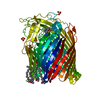
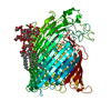
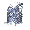
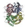
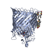
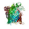
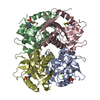
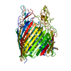
| Title | Crystal structure of the siderophore receptor PiuA from Acinetobacter baumannii | ||||||
 Components Components | TONB-DEPENDENT SIDEROPHORE RECEPTOR | ||||||
 Keywords Keywords | METAL TRANSPORT / SIDEROPHORE RECEPTOR / OUTER-MEMBRANE PROTEIN | ||||||
| Function / homology |  Function and homology information Function and homology informationsiderophore uptake transmembrane transporter activity / porin activity / pore complex / cell outer membrane / signaling receptor activity Similarity search - Function | ||||||
| Biological species |  ACINETOBACTER BAUMANNII (bacteria) ACINETOBACTER BAUMANNII (bacteria) | ||||||
| Method |  X-RAY DIFFRACTION / SYNCHROTRON / MOLECULAR REPLACEMENT / Resolution: 1.94 Å X-RAY DIFFRACTION / SYNCHROTRON / MOLECULAR REPLACEMENT / Resolution: 1.94 Å | ||||||
 Authors Authors | Moynie, L. / Tortajada, A. / Naismith, J.H. | ||||||
 Citation Citation | Journal: Antimicrob. Agents Chemother. / Year: 2017 Title: Structure and Function of the PiuA and PirA Siderophore-Drug Receptors from Pseudomonas aeruginosa and Acinetobacter baumannii. Authors: Moynie, L. / Luscher, A. / Rolo, D. / Pletzer, D. / Tortajada, A. / Weingart, H. / Braun, Y. / Page, M.G. / Naismith, J.H. / Kohler, T. | ||||||
| History |
|
- Structure visualization
| Structure viewer | Molecule: MolmilJmol/JSmol |
|---|
- Downloads & links
-Download
| PDBx/mmCIF format | 5fp1.cif.gz | 169 KB | Display | PDBx/mmCIF format |
|---|---|---|---|---|
| PDB format | pdb5fp1.ent.gz | 132.2 KB | Display | PDB format |
| PDBx/mmJSON format | 5fp1.json.gz | Tree view | PDBx/mmJSON format | |
| Others |  Other downloads Other downloads |
-Validation report
| Arichive directory | https://data.pdbj.org/pub/pdb/validation_reports/fp/5fp1ftp://data.pdbj.org/pub/pdb/validation_reports/fp/5fp1 | HTTPS FTP |
|---|
-Related structure data
| Related structure data |  5fokSC  5fp2C  5fr8C S: Starting model for refinement C: citing same article ( |
|---|---|
| Similar structure data |
-Links
 PDBj
PDBj- Assembly
| Deposited unit | 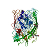
| ||||||||
|---|---|---|---|---|---|---|---|---|---|
| 1 |
| ||||||||
| Unit cell |
|
-Components
| #1: Protein | Mass: 78665.672 Da / Num. of mol.: 1 / Fragment: UNP RESIDUES 29-743 Source method: isolated from a genetically manipulated source Source: (gene. exp.) ACINETOBACTER BAUMANNII (bacteria) / Plasmid: PTAMAHISTEV / Production host: | ||||
|---|---|---|---|---|---|
| #2: Chemical | ChemComp-PO4 /   Mass: 94.971 Da / Num. of mol.: 1 / Source method: obtained synthetically / Formula: PO4 Mass: 94.971 Da / Num. of mol.: 1 / Source method: obtained synthetically / Formula: PO4 | ||||
| #3: Chemical | ChemComp-C8E / (   Mass: 306.438 Da / Num. of mol.: 8 / Source method: obtained synthetically / Formula: C16H34O5 / Comment: C8E, detergent*YM Mass: 306.438 Da / Num. of mol.: 8 / Source method: obtained synthetically / Formula: C16H34O5 / Comment: C8E, detergent*YM#4: Water | ChemComp-HOH / |  Mass: 18.015 Da / Num. of mol.: 792 / Source method: isolated from a natural source / Formula: H2O Mass: 18.015 Da / Num. of mol.: 792 / Source method: isolated from a natural source / Formula: H2OHas protein modification | Y | |
-Experimental details
-Experiment
| Experiment | Method: X-RAY DIFFRACTION / Number of used crystals: 1 |
|---|
- Sample preparation
| Crystal | Density Matthews: 2.2 Å3/Da / Density % sol: 45 % / Description: NONE |
|---|---|
| Crystal grow | pH: 4.6 Details: 1 M DIAMMONIUM HYDROGEN PHOSPHATE, 0.1 M SODIUM ACETATE PH 4.6 |
-Data collection
| Diffraction | Mean temperature: 100 K |
|---|---|
| Diffraction source | Source: SYNCHROTRON / Site: Diamond  / Beamline: I04 / Wavelength: 0.91741 / Beamline: I04 / Wavelength: 0.91741 |
| Detector | Type: DECTRIS PILATUS 6M / Detector: PIXEL / Date: Feb 20, 2015 |
| Radiation | Protocol: SINGLE WAVELENGTH / Monochromatic (M) / Laue (L): M / Scattering type: x-ray |
| Radiation wavelength | Wavelength: 0.91741 Å / Relative weight: 1 |
| Reflection | Resolution: 1.94→65.29 Å / Num. obs: 104869 / % possible obs: 99.7 % / Observed criterion σ(I): 2 / Redundancy: 7.4 % / Rmerge(I) obs: 0.09 / Net I/σ(I): 16.6 |
| Reflection shell | Resolution: 1.94→1.99 Å / Redundancy: 7.5 % / Rmerge(I) obs: 0.66 / Mean I/σ(I) obs: 3 / % possible all: 99.4 |
- Processing
| Software |
| ||||||||||||||||||||||||||||||||||||||||||||||||||||||||||||||||||||||||||||||||||||||||||||||||||||||||||||||||||||||||||||||||||||||||||||||||||||||||||||||||||||||||||||||||||||||
|---|---|---|---|---|---|---|---|---|---|---|---|---|---|---|---|---|---|---|---|---|---|---|---|---|---|---|---|---|---|---|---|---|---|---|---|---|---|---|---|---|---|---|---|---|---|---|---|---|---|---|---|---|---|---|---|---|---|---|---|---|---|---|---|---|---|---|---|---|---|---|---|---|---|---|---|---|---|---|---|---|---|---|---|---|---|---|---|---|---|---|---|---|---|---|---|---|---|---|---|---|---|---|---|---|---|---|---|---|---|---|---|---|---|---|---|---|---|---|---|---|---|---|---|---|---|---|---|---|---|---|---|---|---|---|---|---|---|---|---|---|---|---|---|---|---|---|---|---|---|---|---|---|---|---|---|---|---|---|---|---|---|---|---|---|---|---|---|---|---|---|---|---|---|---|---|---|---|---|---|---|---|---|---|
| Refinement | Method to determine structure: MOLECULAR REPLACEMENT Starting model: PDB ENTRY 5FOK Resolution: 1.94→65.29 Å / Cor.coef. Fo:Fc: 0.959 / Cor.coef. Fo:Fc free: 0.952 / SU B: 1.854 / SU ML: 0.054 / Cross valid method: THROUGHOUT / ESU R: 0.091 / ESU R Free: 0.09 / Stereochemistry target values: MAXIMUM LIKELIHOOD Details: HYDROGENS HAVE BEEN ADDED IN THE RIDING POSITIONS. U VALUES REFINED INDIVIDUALLY
| ||||||||||||||||||||||||||||||||||||||||||||||||||||||||||||||||||||||||||||||||||||||||||||||||||||||||||||||||||||||||||||||||||||||||||||||||||||||||||||||||||||||||||||||||||||||
| Solvent computation | Ion probe radii: 0.8 Å / Shrinkage radii: 0.8 Å / VDW probe radii: 1.2 Å / Solvent model: BABINET MODEL WITH MASK | ||||||||||||||||||||||||||||||||||||||||||||||||||||||||||||||||||||||||||||||||||||||||||||||||||||||||||||||||||||||||||||||||||||||||||||||||||||||||||||||||||||||||||||||||||||||
| Displacement parameters | Biso mean: 28.297 Å2
| ||||||||||||||||||||||||||||||||||||||||||||||||||||||||||||||||||||||||||||||||||||||||||||||||||||||||||||||||||||||||||||||||||||||||||||||||||||||||||||||||||||||||||||||||||||||
| Refinement step | Cycle: LAST / Resolution: 1.94→65.29 Å
| ||||||||||||||||||||||||||||||||||||||||||||||||||||||||||||||||||||||||||||||||||||||||||||||||||||||||||||||||||||||||||||||||||||||||||||||||||||||||||||||||||||||||||||||||||||||
| Refine LS restraints |
|