[English] 日本語
Yorodumi
- PDB-6i98: Structure of the ferrioxamine B transporter FoxA from Pseudomonas... -

+
Open data


ID or keywords:

Loading...

-
Basic information

Entry
Database: PDB / ID: 6i98
TitleStructure of the ferrioxamine B transporter FoxA from Pseudomonas aeruginosa, apo state
ComponentsFerric hydroxamate uptake
KeywordsMEMBRANE PROTEIN
Function / homology
Function and homology information


siderophore transport / siderophore uptake transmembrane transporter activity / outer membrane / cell outer membrane / signaling receptor activity / cell surface receptor signaling pathway
Similarity search - Function
Secretin/TonB, short N-terminal domain / Secretin and TonB N terminus short domain / TonB-dependent siderophore receptor / TonB-dependent receptor (TBDR) proteins profile. / Vitamin B12 transporter BtuB-like / TonB-dependent receptor-like, beta-barrel / TonB dependent receptor-like, beta-barrel / TonB-dependent receptor, plug domain superfamily / TonB-dependent receptor, plug domain / TonB-dependent receptor-like, beta-barrel domain superfamily / TonB-dependent Receptor Plug Domain
Similarity search - Domain/homology
3-HYDROXY-TETRADECANOIC ACID / NICKEL (II) ION / Ferrichrome-iron receptor / Ferrioxamine receptor FoxA
Similarity search - Component
Biological speciesPseudomonas aeruginosa (bacteria)
MethodX-RAY DIFFRACTION / SYNCHROTRON / MOLECULAR REPLACEMENT / molecular replacement / Resolution: 2.8 Å
AuthorsJosts, I. / Tidow, H.
Funding support Germany, 1items
OrganizationGrant numberCountry
German Research Foundation Germany
Citation
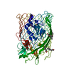
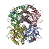
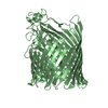
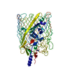
Journal: Elife / Year: 2019
Title: Ternary structure of the outer membrane transporter FoxA with resolved signalling domain provides insights into TonB-mediated siderophore uptake.
Authors: Josts, I. / Veith, K. / Tidow, H.
History
DepositionNov 22, 2018Deposition site: PDBE / Processing site: PDBE
Revision 1.0Aug 28, 2019Provider: repository / Type: Initial release

-
Structure visualization

Structure viewerMolecule:
MolmilJmol/JSmol

Downloads & links

-
Assembly

Deposited unit
A: Ferric hydroxamate uptake
hetero molecules


Theoretical massNumber of molelcules
Total (without water)79,00727
Polymers75,4221
Non-polymers3,58526
Water25214
1


  • Idetical with deposited unit
  • defined by author&software
TypeNameSymmetry operationNumber
identity operation1_555x,y,z1
Buried area6430 Å2
ΔGint-153 kcal/mol
Surface area27050 Å2
MethodPISA
Unit cell
Length a, b, c (Å)174.554, 174.554, 180.245
Angle α, β, γ (deg.)90.000, 90.000, 120.000
Int Tables number182
Space group name H-MP6322

-
Components

-
Protein , 1 types, 1 molecules A

#1: Protein Ferric hydroxamate uptake / Ferrichrome-iron receptor / Ferrioxamine receptor FoxA / TonB-dependent siderophore receptor


Mass: 75421.867 Da / Num. of mol.: 1
Source method: isolated from a genetically manipulated source
Source: (gene. exp.) Pseudomonas aeruginosa (bacteria)
Gene: fhuA, fhuA_5, C0044_18480, CAZ10_13360, CCBH4851_00003, DZ940_30305, EGY23_22030, PAERUG_E15_London_28_01_14_03466
Production host: Escherichia coli (E. coli) / References: UniProt: A0A0C7CQY7, UniProt: Q9I116*PLUS

-
Non-polymers , 7 types, 40 molecules

#2: Chemical ChemComp-NI / NICKEL (II) ION


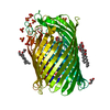
Mass: 58.693 Da / Num. of mol.: 1 / Source method: obtained synthetically / Formula: Ni
#3: Chemical
ChemComp-GOL / GLYCEROL / GLYCERIN / PROPANE-1,2,3-TRIOL


Mass: 92.094 Da / Num. of mol.: 4 / Source method: obtained synthetically / Formula: C3H8O3
#4: Chemical
ChemComp-C8E / (HYDROXYETHYLOXY)TRI(ETHYLOXY)OCTANE


Mass: 306.438 Da / Num. of mol.: 4 / Source method: obtained synthetically / Formula: C16H34O5 / Comment: C8E, detergent*YM
#5: Chemical ChemComp-FTT / 3-HYDROXY-TETRADECANOIC ACID / 3-HYDROXY-MYRISTIC ACID


Mass: 244.370 Da / Num. of mol.: 3 / Source method: obtained synthetically / Formula: C14H28O3
#6: Chemical
ChemComp-SO4 / SULFATE ION


Mass: 96.063 Da / Num. of mol.: 12 / Source method: isolated from a natural source / Formula: SO4
#7: Chemical ChemComp-NA / SODIUM ION


Mass: 22.990 Da / Num. of mol.: 2 / Source method: obtained synthetically / Formula: Na
#8: Water ChemComp-HOH / water


Mass: 18.015 Da / Num. of mol.: 14 / Source method: isolated from a natural source / Formula: H2O

-
Experimental details

-
Experiment

ExperimentMethod: X-RAY DIFFRACTION / Number of used crystals: 1

-
Sample preparation

CrystalDensity Matthews: 5.24 Å3/Da / Density % sol: 76.52 %
Crystal growTemperature: 295 K / Method: vapor diffusion, sitting drop / Details: 1.8 M Ammonium sulfate, 0.1M HEPES pH 7.4

-
Data collection

DiffractionMean temperature: 100 K / Serial crystal experiment: N
Diffraction sourceSource: SYNCHROTRON / Site: PETRA III, EMBL c/o DESY / Beamline: P13 (MX1) / Wavelength: 1.03332 Å
DetectorType: DECTRIS PILATUS 300K / Detector: PIXEL / Date: Jun 5, 2017
RadiationProtocol: SINGLE WAVELENGTH / Monochromatic (M) / Laue (L): M / Scattering type: x-ray
Radiation wavelengthWavelength: 1.03332 Å / Relative weight: 1
ReflectionResolution: 2.8→48.53 Å / Num. obs: 40394 / % possible obs: 99.9 % / Redundancy: 10.7 % / CC1/2: 0.997 / Rmerge(I) obs: 0.206 / Rpim(I) all: 0.092 / Rrim(I) all: 0.226 / Net I/σ(I): 10
Reflection shellResolution: 2.8→2.91 Å / Mean I/σ(I) obs: 1.6 / Num. unique obs: 4492 / CC1/2: 0.547 / % possible all: 100

-
Phasing

PhasingMethod: molecular replacement

-
Processing

Software
NameVersionClassificationNB
REFMAC5.8.0158refinement
XDSdata reduction
Aimlessdata scaling
PHASERphasing
PDB_EXTRACT3.24data extraction
RefinementMethod to determine structure: MOLECULAR REPLACEMENT / Resolution: 2.8→151.17 Å / Cor.coef. Fo:Fc: 0.92 / Cor.coef. Fo:Fc free: 0.892 / SU B: 24.424 / SU ML: 0.216 / Cross valid method: THROUGHOUT / σ(F): 0 / ESU R: 0.355 / ESU R Free: 0.281 / Stereochemistry target values: MAXIMUM LIKELIHOOD
Details: HYDROGENS HAVE BEEN ADDED IN THE RIDING POSITIONS U VALUES : WITH TLS ADDED
RfactorNum. reflection% reflectionSelection details
Rfree0.2599 2028 5 %RANDOM
Rwork0.2133 ---
obs0.2156 38331 99.84 %-
Solvent computationIon probe radii: 0.8 Å / Shrinkage radii: 0.8 Å / VDW probe radii: 1.2 Å / Solvent model: MASK
Displacement parametersBiso max: 141 Å2 / Biso mean: 58.049 Å2 / Biso min: 30 Å2
Baniso -1Baniso -2Baniso -3
1-0.56 Å20.28 Å20 Å2
2--0.56 Å20 Å2
3----1.81 Å2
Refinement stepCycle: final / Resolution: 2.8→151.17 Å
ProteinNucleic acidLigandSolventTotal
Num. atoms5325 0 193 14 5532
Biso mean--92.28 50.97 -
Num. residues----677
Refine LS restraints
Refine-IDTypeDev idealDev ideal targetNumber
X-RAY DIFFRACTIONr_bond_refined_d0.0210.0195619
X-RAY DIFFRACTIONr_bond_other_d0.0020.024956
X-RAY DIFFRACTIONr_angle_refined_deg2.341.977588
X-RAY DIFFRACTIONr_angle_other_deg1.151311522
X-RAY DIFFRACTIONr_dihedral_angle_1_deg8.8485676
X-RAY DIFFRACTIONr_dihedral_angle_2_deg39.72924.143280
X-RAY DIFFRACTIONr_dihedral_angle_3_deg19.6915858
X-RAY DIFFRACTIONr_dihedral_angle_4_deg20.751535
X-RAY DIFFRACTIONr_chiral_restr0.1290.2801
X-RAY DIFFRACTIONr_gen_planes_refined0.010.026253
X-RAY DIFFRACTIONr_gen_planes_other0.0020.021186
LS refinement shellResolution: 2.8→2.873 Å / Rfactor Rfree error: 0 / Total num. of bins used: 20
RfactorNum. reflection% reflection
Rfree0.372 155 -
Rwork0.328 2769 -
all-2924 -
obs--99.97 %
Refinement TLS params.Method: refined / Origin x: -6.7742 Å / Origin y: -70.3826 Å / Origin z: -12.8859 Å
111213212223313233
T0.0501 Å2-0.0038 Å20.055 Å2-0.0095 Å2-0.0236 Å2--0.1555 Å2
L0.4411 °2-0.0844 °2-0.0222 °2-0.4744 °2-0.0385 °2--0.4213 °2
S0.023 Å °-0.0574 Å °0.0847 Å °-0.0016 Å °-0.0017 Å °-0.0176 Å °-0.1202 Å °-0.0082 Å °-0.0213 Å °

+
About Yorodumi

-
News

-
Feb 9, 2022. New format data for meta-information of EMDB entries

New format data for meta-information of EMDB entries

  • Version 3 of the EMDB header file is now the official format.
  • The previous official version 1.9 will be removed from the archive.

Related info.:EMDB header

External links:wwPDB to switch to version 3 of the EMDB data model

-
Aug 12, 2020. Covid-19 info

Covid-19 info

URL: https://pdbjlvh1.pdbj.org/emnavi/covid19.php

New page: Covid-19 featured information page in EM Navigator.

Related info.:Covid-19 info / Mar 5, 2020. Novel coronavirus structure data

+
Mar 5, 2020. Novel coronavirus structure data

Novel coronavirus structure data

Related info.:Yorodumi Speices / Aug 12, 2020. Covid-19 info

External links:COVID-19 featured content - PDBj / Molecule of the Month (242):Coronavirus Proteases

+
Jan 31, 2019. EMDB accession codes are about to change! (news from PDBe EMDB page)

EMDB accession codes are about to change! (news from PDBe EMDB page)

  • The allocation of 4 digits for EMDB accession codes will soon come to an end. Whilst these codes will remain in use, new EMDB accession codes will include an additional digit and will expand incrementally as the available range of codes is exhausted. The current 4-digit format prefixed with “EMD-” (i.e. EMD-XXXX) will advance to a 5-digit format (i.e. EMD-XXXXX), and so on. It is currently estimated that the 4-digit codes will be depleted around Spring 2019, at which point the 5-digit format will come into force.
  • The EM Navigator/Yorodumi systems omit the EMD- prefix.

Related info.:Q: What is EMD? / ID/Accession-code notation in Yorodumi/EM Navigator

External links:EMDB Accession Codes are Changing Soon! / Contact to PDBj

+
Jul 12, 2017. Major update of PDB

Major update of PDB

  • wwPDB released updated PDB data conforming to the new PDBx/mmCIF dictionary.
  • This is a major update changing the version number from 4 to 5, and with Remediation, in which all the entries are updated.
  • In this update, many items about electron microscopy experimental information are reorganized (e.g. em_software).
  • Now, EM Navigator and Yorodumi are based on the updated data.

External links:wwPDB Remediation / Enriched Model Files Conforming to OneDep Data Standards Now Available in the PDB FTP Archive

-
Yorodumi

Thousand views of thousand structures

  • Yorodumi is a browser for structure data from EMDB, PDB, SASBDB, etc.
  • This page is also the successor to EM Navigator detail page, and also detail information page/front-end page for Omokage search.
  • The word "yorodu" (or yorozu) is an old Japanese word meaning "ten thousand". "mi" (miru) is to see.

Related info.:EMDB / PDB / SASBDB / Comparison of 3 databanks / Yorodumi Search / Aug 31, 2016. New EM Navigator & Yorodumi / Yorodumi Papers / Jmol/JSmol / Function and homology information / Changes in new EM Navigator and Yorodumi

Read more