transferase/DNA / NUCLEOTIDYLTRANSFERASE / DNA REPAIR / DNA MISMATCH / BASE EXCISION REPAIR / transferase-DNA COMPLEX
Function / homology
Function and homology information
Resolution of AP sites via the single-nucleotide replacement pathway / immunoglobulin heavy chain V-D-J recombination / Resolution of AP sites via the multiple-nucleotide patch replacement pathway / Abasic sugar-phosphate removal via the single-nucleotide replacement pathway / APEX1-Independent Resolution of AP Sites via the Single Nucleotide Replacement Pathway / Lyases; Carbon-oxygen lyases; Other carbon-oxygen lyases / homeostasis of number of cells / pyrimidine dimer repair / POLB-Dependent Long Patch Base Excision Repair / PCNA-Dependent Long Patch Base Excision Repair ...Resolution of AP sites via the single-nucleotide replacement pathway / immunoglobulin heavy chain V-D-J recombination / Resolution of AP sites via the multiple-nucleotide patch replacement pathway / Abasic sugar-phosphate removal via the single-nucleotide replacement pathway / APEX1-Independent Resolution of AP Sites via the Single Nucleotide Replacement Pathway / Lyases; Carbon-oxygen lyases; Other carbon-oxygen lyases / homeostasis of number of cells / pyrimidine dimer repair / POLB-Dependent Long Patch Base Excision Repair / PCNA-Dependent Long Patch Base Excision Repair / 5'-deoxyribose-5-phosphate lyase activity / response to hyperoxia / lymph node development / spleen development / salivary gland morphogenesis / somatic hypermutation of immunoglobulin genes / base-excision repair, gap-filling / DNA-(apurinic or apyrimidinic site) endonuclease activity / class I DNA-(apurinic or apyrimidinic site) endonuclease activity / DNA-(apurinic or apyrimidinic site) lyase / response to gamma radiation / spindle microtubule / base-excision repair / DNA-templated DNA replication / double-strand break repair via nonhomologous end joining / intrinsic apoptotic signaling pathway in response to DNA damage / neuron apoptotic process / in utero embryonic development / DNA-directed DNA polymerase / microtubule binding / damaged DNA binding / microtubule / response to ethanol / DNA-directed DNA polymerase activity / lyase activity / Ub-specific processing proteases / inflammatory response / DNA repair / DNA damage response / enzyme binding / protein-containing complex / nucleoplasm / metal ion binding / nucleus / cytoplasm Similarity search - Function
Beta Polymerase; domain 3 / DNA polymerase, thumb domain / DNA polymerase beta, N-terminal domain-like / DNA polymerase family X, beta-like / DNA polymerase beta, palm domain / DNA polymerase beta palm / DNA polymerase lambda, fingers domain / Fingers domain of DNA polymerase lambda / DNA-directed DNA polymerase X / DNA polymerase X family ...Beta Polymerase; domain 3 / DNA polymerase, thumb domain / DNA polymerase beta, N-terminal domain-like / DNA polymerase family X, beta-like / DNA polymerase beta, palm domain / DNA polymerase beta palm / DNA polymerase lambda, fingers domain / Fingers domain of DNA polymerase lambda / DNA-directed DNA polymerase X / DNA polymerase X family / DNA polymerase family X, binding site / DNA polymerase family X signature. / DNA polymerase beta-like, N-terminal domain / DNA polymerase lambda lyase domain superfamily / Helix-hairpin-helix domain / DNA polymerase family X / DNA polymerase beta, thumb domain / DNA polymerase, thumb domain superfamily / DNA polymerase beta thumb / Beta Polymerase, domain 2 / Helix-hairpin-helix DNA-binding motif, class 1 / Helix-hairpin-helix DNA-binding motif class 1 / Beta Polymerase; domain 2 / Nucleotidyltransferase superfamily / 5' to 3' exonuclease, C-terminal subdomain / DNA polymerase; domain 1 / 2-Layer Sandwich / Orthogonal Bundle / Mainly Alpha / Alpha Beta Similarity search - Domain/homology
Mass: 4844.145 Da / Num. of mol.: 1 / Source method: obtained synthetically
#2: DNA chain
5'-D(*GP*CP*TP*GP*AP*TP*GP*CP*GP*CP*C)-3'
Mass: 3350.185 Da / Num. of mol.: 1 / Source method: obtained synthetically
#3: DNA chain
5'-D(P*GP*TP*CP*GP*G)-3'
Mass: 1536.035 Da / Num. of mol.: 1 / Source method: obtained synthetically
-
Protein , 1 types, 1 molecules A
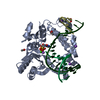
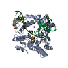
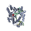
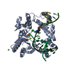
#4: Protein
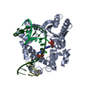
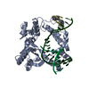
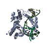
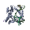
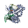
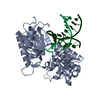
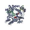
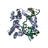
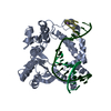
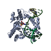
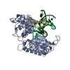
DNApolymerasebeta
Mass: 38241.672 Da / Num. of mol.: 1 Source method: isolated from a genetically manipulated source Source: (gene. exp.) Homo sapiens (human) / Gene: POLB / Production host: Escherichia coli (E. coli) / References: UniProt: P06746, DNA-directed DNA polymerase
Type: RIGAKU RAXIS IV / Detector: IMAGE PLATE / Date: Oct 1, 1998 / Details: YALE MIRRORS
Radiation
Monochromator: YALE MIRRORS / Protocol: SINGLE WAVELENGTH / Monochromatic (M) / Laue (L): M / Scattering type: x-ray
Radiation wavelength
Wavelength: 1.5418 Å / Relative weight: 1
Reflection
Resolution: 2.6→50 Å / Num. obs: 13572 / % possible obs: 98 % / Observed criterion σ(I): -3 / Redundancy: 4 % / Rmerge(I) obs: 0.086 / Net I/σ(I): 13.8
Reflection shell
Resolution: 2.6→2.69 Å / Redundancy: 3.6 % / Rmerge(I) obs: 0.262 / Mean I/σ(I) obs: 4.1 / % possible all: 95.8
-
Processing
Software
Name
Classification
DENZO
datareduction
SCALEPACK
datascaling
CNS
refinement
CNS
phasing
Refinement
Method to determine structure: MOLECULAR REPLACEMENT / Resolution: 2.6→50 Å / Isotropic thermal model: ANISOTROPIC / Cross valid method: THROUGHOUT / σ(F): 0 / Stereochemistry target values: ENGH & HUBER Details: CNS BULK SOLVENT MODEL. PARAMETERS FOR B-FORM SPECIFIC DNA DIHEDRAL RESTRAINTS IN CNS WERE EXCLUDED BECAUSE MANY OF THE NUCLEIC ACIDS DEVIATE FROM STRICT B-FORM GEOMETRY.
In the structure databanks used in Yorodumi, some data are registered as the other names, "COVID-19 virus" and "2019-nCoV". Here are the details of the virus and the list of structure data.
Jan 31, 2019. EMDB accession codes are about to change! (news from PDBe EMDB page)
EMDB accession codes are about to change! (news from PDBe EMDB page)
The allocation of 4 digits for EMDB accession codes will soon come to an end. Whilst these codes will remain in use, new EMDB accession codes will include an additional digit and will expand incrementally as the available range of codes is exhausted. The current 4-digit format prefixed with “EMD-” (i.e. EMD-XXXX) will advance to a 5-digit format (i.e. EMD-XXXXX), and so on. It is currently estimated that the 4-digit codes will be depleted around Spring 2019, at which point the 5-digit format will come into force.
The EM Navigator/Yorodumi systems omit the EMD- prefix.
Related info.:Q: What is EMD? / ID/Accession-code notation in Yorodumi/EM Navigator
Yorodumi is a browser for structure data from EMDB, PDB, SASBDB, etc.
This page is also the successor to EM Navigator detail page, and also detail information page/front-end page for Omokage search.
The word "yorodu" (or yorozu) is an old Japanese word meaning "ten thousand". "mi" (miru) is to see.
Related info.:EMDB / PDB / SASBDB / Comparison of 3 databanks / Yorodumi Search / Aug 31, 2016. New EM Navigator & Yorodumi / Yorodumi Papers / Jmol/JSmol / Function and homology information / Changes in new EM Navigator and Yorodumi
 Movie
Movie Controller

 Yorodumi
 Open data
 Basic information
 Components
 Keywords
 Function and homology information
 Homo sapiens (human)
 X-RAY DIFFRACTION /
  Authors
 Citation
 Structure visualization
 Downloads & links
 Other downloads


 PDBj
PDBj


 Assembly
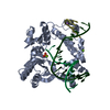


 Mass: 24.305 Da / Num. of mol.: 2 / Source method: obtained synthetically / Formula: Mg
 Mass: 22.990 Da / Num. of mol.: 2 / Source method: obtained synthetically / Formula: Na
 Mass: 94.971 Da / Num. of mol.: 1 / Source method: obtained synthetically / Formula: PO4
 Sample preparation
 Processing