 Movie
Movie Controller

[English] 日本語
 Yorodumi
- PDB-1uwx: P1.2 serosubtype antigen derived from N. meningitidis PorA in com... -
+ Open data

- Basic information
| Entry | Database: PDB / ID: 1uwx | ||||||
|---|---|---|---|---|---|---|---|
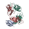
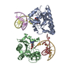
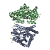
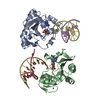
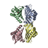
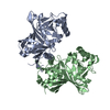
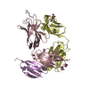
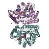
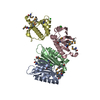
| Title | P1.2 serosubtype antigen derived from N. meningitidis PorA in complex with Fab fragment | ||||||
 Components Components |
| ||||||
 Keywords Keywords | IMMUNE SYSTEM / ANTIBODY-COMPLEX / FAB / IMMUNOGLOBULIN / PROTEIN G / PORA / ANTIBODY / IGB | ||||||
| Function / homology |  Function and homology information Function and homology informationalpha-beta T cell receptor complex / IgG binding / IgG immunoglobulin complex / porin activity / pore complex / B cell differentiation / cell outer membrane / monoatomic ion transmembrane transport / adaptive immune response / extracellular region / plasma membrane Similarity search - Function | ||||||
| Biological species |  STREPTOCOCCUS SP. (bacteria) STREPTOCOCCUS SP. (bacteria) NEISSERIA MENINGITIDIS (bacteria) NEISSERIA MENINGITIDIS (bacteria) | ||||||
| Method |  X-RAY DIFFRACTION / SYNCHROTRON / MOLECULAR REPLACEMENT / Resolution: 2.2 Å X-RAY DIFFRACTION / SYNCHROTRON / MOLECULAR REPLACEMENT / Resolution: 2.2 Å | ||||||
 Authors Authors | Tzitzilonis, C. / Prince, S.M. / Collins, R.F. / Maiden, M.C.J. / Feavers, I.M. / Derrick, J.P. | ||||||
 Citation Citation | Journal: Proteins: Struct., Funct., Bioinf. / Year: 2005 Title: Structural Variation and Immune Recognition of the P1.2 Subtype Meningococcal Antigen. Authors: Tzitzilonis, C. / Prince, S.M. / Collins, R.F. / Achtman, M. / Feavers, I.M. / Maiden, M.C.J. / Derrick, J.P. | ||||||
| History |
| ||||||
| Remark 700 | SHEET THE SHEET STRUCTURE OF THIS MOLECULE IS BIFURCATED. IN ORDER TO REPRESENT THIS FEATURE IN ... SHEET THE SHEET STRUCTURE OF THIS MOLECULE IS BIFURCATED. IN ORDER TO REPRESENT THIS FEATURE IN THE SHEET RECORDS BELOW, TWO SHEETS ARE DEFINED. |
- Structure visualization
| Structure viewer | Molecule: MolmilJmol/JSmol |
|---|
- Downloads & links
-Download
| PDBx/mmCIF format | 1uwx.cif.gz | 207.8 KB | Display | PDBx/mmCIF format |
|---|---|---|---|---|
| PDB format | pdb1uwx.ent.gz | 166.5 KB | Display | PDB format |
| PDBx/mmJSON format | 1uwx.json.gz | Tree view | PDBx/mmJSON format | |
| Others |  Other downloads Other downloads |
-Validation report
| Arichive directory | https://data.pdbj.org/pub/pdb/validation_reports/uw/1uwxftp://data.pdbj.org/pub/pdb/validation_reports/uw/1uwx | HTTPS FTP |
|---|
-Related structure data
| Related structure data |  1qkzS S: Starting model for refinement |
|---|---|
| Similar structure data |

-Links
 PDBj
PDBj


- Assembly
| Deposited unit | 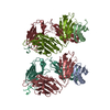
| |||||||||||||||||||||||||||||||||||||||||||||||||||||||||||||||||||||||||||||||
|---|---|---|---|---|---|---|---|---|---|---|---|---|---|---|---|---|---|---|---|---|---|---|---|---|---|---|---|---|---|---|---|---|---|---|---|---|---|---|---|---|---|---|---|---|---|---|---|---|---|---|---|---|---|---|---|---|---|---|---|---|---|---|---|---|---|---|---|---|---|---|---|---|---|---|---|---|---|---|---|---|
| 1 | 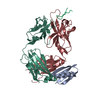
| |||||||||||||||||||||||||||||||||||||||||||||||||||||||||||||||||||||||||||||||
| 2 | 
| |||||||||||||||||||||||||||||||||||||||||||||||||||||||||||||||||||||||||||||||
| Unit cell |
| |||||||||||||||||||||||||||||||||||||||||||||||||||||||||||||||||||||||||||||||
| Noncrystallographic symmetry (NCS) | NCS domain:
NCS domain segments:
NCS ensembles :
|
-Components
| #1: Protein | Mass: 6881.566 Da / Num. of mol.: 2 / Fragment: RESIDUES 56-118 (DOMAIN II) Source method: isolated from a genetically manipulated source Source: (gene. exp.) STREPTOCOCCUS SP. (bacteria) / Description: HYBRIDOMA / Production host: #2: Antibody | Mass: 24522.346 Da / Num. of mol.: 2 / Fragment: RESIDUES 1-215 (FAB FRAGMENT, HEAVY CHAIN) / Source method: isolated from a natural source / Source: (natural) #3: Antibody | Mass: 23218.730 Da / Num. of mol.: 2 / Fragment: RESIDUES 3-214 (FAB FRAGMENT, LIGHT CHAIN) / Source method: isolated from a natural source / Details: HYBRIDOMA / Source: (natural) #4: Protein/peptide | Mass: 1512.706 Da / Num. of mol.: 2 / Fragment: RESIDUES 16-28 / Source method: obtained synthetically Details: RAISED AGAINST THE P1.2 SEROSUBTYPE ANTIGEN SEQUENCE FROM THE PORA PROTEIN FROM NEISSERI MENINGITIDIS Source: (synth.) NEISSERIA MENINGITIDIS (bacteria) / References: UniProt: Q51220, UniProt: Q9JPT2*PLUS#5: Water | ChemComp-HOH / |  Mass: 18.015 Da / Num. of mol.: 395 / Source method: isolated from a natural source / Formula: H2O Mass: 18.015 Da / Num. of mol.: 395 / Source method: isolated from a natural source / Formula: H2OHas protein modification | Y | |
|---|
-Experimental details
-Experiment
| Experiment | Method: X-RAY DIFFRACTION / Number of used crystals: 2 |
|---|
- Sample preparation
| Crystal | Density Matthews: 2.7 Å3/Da / Density % sol: 55.78 % |
|---|---|
| Crystal grow | pH: 5 / Details: pH 5.00 |
-Data collection
| Diffraction | Mean temperature: 100 K |
|---|---|
| Diffraction source | Source: SYNCHROTRON / Site: SRS  / Beamline: PX9.6 / Wavelength: 0.87 / Beamline: PX9.6 / Wavelength: 0.87 |
| Detector | Type: ADSC CCD / Detector: CCD |
| Radiation | Protocol: SINGLE WAVELENGTH / Monochromatic (M) / Laue (L): M / Scattering type: x-ray |
| Radiation wavelength | Wavelength: 0.87 Å / Relative weight: 1 |
| Reflection | Resolution: 2.2→20 Å / Num. obs: 50717 / % possible obs: 87 % / Redundancy: 5.1 % / Rmerge(I) obs: 0.073 / Net I/σ(I): 11.7 |
| Reflection shell | Resolution: 2.2→2.26 Å / Redundancy: 1.3 % / Rmerge(I) obs: 0.36 / Mean I/σ(I) obs: 1.8 / % possible all: 45 |
- Processing
| Software |
| ||||||||||||||||||||||||||||||||||||||||||||||||||||||||||||||||||||||||||||||||||||||||||||||||||||||||||||||||||||||||||||||||||||||||||||||||||||||||||||||||||||||||||||||||||||||
|---|---|---|---|---|---|---|---|---|---|---|---|---|---|---|---|---|---|---|---|---|---|---|---|---|---|---|---|---|---|---|---|---|---|---|---|---|---|---|---|---|---|---|---|---|---|---|---|---|---|---|---|---|---|---|---|---|---|---|---|---|---|---|---|---|---|---|---|---|---|---|---|---|---|---|---|---|---|---|---|---|---|---|---|---|---|---|---|---|---|---|---|---|---|---|---|---|---|---|---|---|---|---|---|---|---|---|---|---|---|---|---|---|---|---|---|---|---|---|---|---|---|---|---|---|---|---|---|---|---|---|---|---|---|---|---|---|---|---|---|---|---|---|---|---|---|---|---|---|---|---|---|---|---|---|---|---|---|---|---|---|---|---|---|---|---|---|---|---|---|---|---|---|---|---|---|---|---|---|---|---|---|---|---|
| Refinement | Method to determine structure: MOLECULAR REPLACEMENT Starting model: PDB ENTRY 1QKZ Resolution: 2.2→20 Å / Cor.coef. Fo:Fc: 0.938 / Cor.coef. Fo:Fc free: 0.913 / SU ML: 0.19 / TLS residual ADP flag: UNVERIFIED / Cross valid method: THROUGHOUT / ESU R: 0.328 / ESU R Free: 0.243 / Stereochemistry target values: MAXIMUM LIKELIHOOD
| ||||||||||||||||||||||||||||||||||||||||||||||||||||||||||||||||||||||||||||||||||||||||||||||||||||||||||||||||||||||||||||||||||||||||||||||||||||||||||||||||||||||||||||||||||||||
| Solvent computation | Ion probe radii: 0.8 Å / Shrinkage radii: 0.8 Å / VDW probe radii: 1.4 Å / Solvent model: BABINET MODEL WITH MASK | ||||||||||||||||||||||||||||||||||||||||||||||||||||||||||||||||||||||||||||||||||||||||||||||||||||||||||||||||||||||||||||||||||||||||||||||||||||||||||||||||||||||||||||||||||||||
| Displacement parameters | Biso mean: 20.41 Å2
| ||||||||||||||||||||||||||||||||||||||||||||||||||||||||||||||||||||||||||||||||||||||||||||||||||||||||||||||||||||||||||||||||||||||||||||||||||||||||||||||||||||||||||||||||||||||
| Refinement step | Cycle: LAST / Resolution: 2.2→20 Å
| ||||||||||||||||||||||||||||||||||||||||||||||||||||||||||||||||||||||||||||||||||||||||||||||||||||||||||||||||||||||||||||||||||||||||||||||||||||||||||||||||||||||||||||||||||||||
| Refine LS restraints |
|