 Movie
Movie Controller

[English] 日本語
 Yorodumi
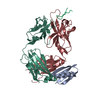
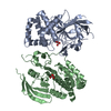
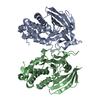
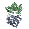
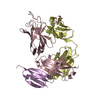
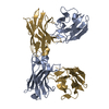
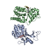
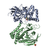
- PDB-1qkz: Fab fragment (MN14C11.6) in complex with a peptide antigen derive... -
+ Open data

- Basic information
| Entry | Database: PDB / ID: 1qkz | ||||||
|---|---|---|---|---|---|---|---|
| Title | Fab fragment (MN14C11.6) in complex with a peptide antigen derived from Neisseria meningitidis P1.7 serosubtype antigen and domain II from Streptococcal protein G | ||||||
 Components Components |
| ||||||
 Keywords Keywords | IMMUNE SYSTEM / FAB / PORA / NEISSERIA MENINGITIDIS / PORIN | ||||||
| Function / homology |  Function and homology information Function and homology informationIgG binding / porin activity / pore complex / cell outer membrane / monoatomic ion transmembrane transport Similarity search - Function | ||||||
| Biological species |  STREPTOCOCCUS SP. (bacteria) STREPTOCOCCUS SP. (bacteria) NEISSERIA MENINGITIDIS (bacteria) NEISSERIA MENINGITIDIS (bacteria) | ||||||
| Method |  X-RAY DIFFRACTION / SYNCHROTRON / MOLECULAR REPLACEMENT / Resolution: 1.95 Å X-RAY DIFFRACTION / SYNCHROTRON / MOLECULAR REPLACEMENT / Resolution: 1.95 Å | ||||||
 Authors Authors | Derrick, J.P. / Feavers, I. / Maiden, M.C.J. | ||||||
 Citation Citation | Journal: J.Mol.Biol. / Year: 1999 Title: Crystal Structure of an Fab Fragment in Complex with a Meningococcal Serosubtype Antigen and a Protein G Domain Authors: Derrick, J.P. / Feavers, I.M. / Maiden, M.C.J. | ||||||
| History |
|
- Structure visualization
| Structure viewer | Molecule: MolmilJmol/JSmol |
|---|
- Downloads & links
-Download
| PDBx/mmCIF format | 1qkz.cif.gz | 119.9 KB | Display | PDBx/mmCIF format |
|---|---|---|---|---|
| PDB format | pdb1qkz.ent.gz | 91.6 KB | Display | PDB format |
| PDBx/mmJSON format | 1qkz.json.gz | Tree view | PDBx/mmJSON format | |
| Others |  Other downloads Other downloads |
-Validation report
| Arichive directory | https://data.pdbj.org/pub/pdb/validation_reports/qk/1qkzftp://data.pdbj.org/pub/pdb/validation_reports/qk/1qkz | HTTPS FTP |
|---|
-Related structure data
| Related structure data | 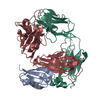 1igcS S: Starting model for refinement |
|---|---|
| Similar structure data |


-Links
 PDBj
PDBj

- Assembly
| Deposited unit | 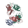
| ||||||||
|---|---|---|---|---|---|---|---|---|---|
| 1 |
| ||||||||
| Unit cell |
|
-Components
| #1: Protein | Mass: 7012.762 Da / Num. of mol.: 1 / Fragment: DOMAIN II / Mutation: YES Source method: isolated from a genetically manipulated source Source: (gene. exp.) STREPTOCOCCUS SP. (bacteria) / Strain: GROUP G / Production host: |
|---|---|
| #2: Antibody | Mass: 23755.592 Da / Num. of mol.: 1 / Fragment: FAB / Source method: isolated from a natural source / Source: (natural) |
| #3: Antibody | Mass: 23954.559 Da / Num. of mol.: 1 / Fragment: FAB / Source method: isolated from a natural source / Source: (natural) |
| #4: Protein/peptide | Mass: 888.946 Da / Num. of mol.: 1 / Fragment: RESIDUES 4-13 / Source method: obtained synthetically / Source: (synth.) NEISSERIA MENINGITIDIS (bacteria) / References: UniProt: Q06140, UniProt: P57041*PLUS |
| #5: Water | ChemComp-HOH /  Mass: 18.015 Da / Num. of mol.: 526 / Source method: isolated from a natural source / Formula: H2O Mass: 18.015 Da / Num. of mol.: 526 / Source method: isolated from a natural source / Formula: H2O |
| Has protein modification | Y |
| Sequence details | THE FAB ANTIBODY STUDIED HERE IS RELATED IN SEQUENCE TO THE SEQUENCES: CHAIN H TO SWS AAD40243 78 ...THE FAB ANTIBODY STUDIED HERE IS RELATED IN SEQUENCE TO THE SEQUENCES: CHAIN H TO SWS AAD40243 78 PERCENT IDENTITY GAMMA1 HEAVY CHAIN OF MAB7 MUS MUSCULUS CHAIN L TO SWS AAD40242 74 PERCENT IDENTITY KAPPA LIGHT CHAIN OF MAB7 MUS MUSCULUS THE RESIDUE NUMBERING SCHEME USED HERE IS THE KABAT NUMBERING |
-Experimental details
-Experiment
| Experiment | Method: X-RAY DIFFRACTION / Number of used crystals: 1 |
|---|
- Sample preparation
| Crystal | Density Matthews: 2.76 Å3/Da / Density % sol: 43.6 % | ||||||||||||||||||||||||||||||
|---|---|---|---|---|---|---|---|---|---|---|---|---|---|---|---|---|---|---|---|---|---|---|---|---|---|---|---|---|---|---|---|
| Crystal grow | pH: 5 / Details: pH 5.00 | ||||||||||||||||||||||||||||||
| Crystal grow | *PLUS Temperature: 293 K / Method: vapor diffusion, hanging drop | ||||||||||||||||||||||||||||||
| Components of the solutions | *PLUS
|
-Data collection
| Diffraction | Mean temperature: 100 K |
|---|---|
| Diffraction source | Source: SYNCHROTRON / Site: SRS  / Beamline: PX9.5 / Wavelength: 1.1 / Beamline: PX9.5 / Wavelength: 1.1 |
| Detector | Type: MARRESEARCH / Detector: IMAGE PLATE |
| Radiation | Protocol: SINGLE WAVELENGTH / Monochromatic (M) / Laue (L): M / Scattering type: x-ray |
| Radiation wavelength | Wavelength: 1.1 Å / Relative weight: 1 |
| Reflection | Resolution: 1.95→30 Å / Num. obs: 34633 / % possible obs: 98.9 % / Observed criterion σ(I): 2 / Redundancy: 5.2 % / Biso Wilson estimate: 13.7 Å2 / Rsym value: 0.037 / Net I/σ(I): 17.2 |
| Reflection shell | Resolution: 1.95→2.04 Å / Rsym value: 0.125 / % possible all: 96 |
| Reflection | *PLUS Num. measured all: 180259 / Rmerge(I) obs: 0.037 |
| Reflection shell | *PLUS % possible obs: 96 % / Rmerge(I) obs: 0.125 |
- Processing
| Software |
| ||||||||||||||||||||||||||||||||||||||||||||||||||||||||||||||||||||||||||||||||||||
|---|---|---|---|---|---|---|---|---|---|---|---|---|---|---|---|---|---|---|---|---|---|---|---|---|---|---|---|---|---|---|---|---|---|---|---|---|---|---|---|---|---|---|---|---|---|---|---|---|---|---|---|---|---|---|---|---|---|---|---|---|---|---|---|---|---|---|---|---|---|---|---|---|---|---|---|---|---|---|---|---|---|---|---|---|---|
| Refinement | Method to determine structure: MOLECULAR REPLACEMENT Starting model: PDB ENTRY 1IGC Resolution: 1.95→10 Å / Cross valid method: THROUGHOUT / σ(F): 0 / ESU R: 0.21
| ||||||||||||||||||||||||||||||||||||||||||||||||||||||||||||||||||||||||||||||||||||
| Refinement step | Cycle: LAST / Resolution: 1.95→10 Å
| ||||||||||||||||||||||||||||||||||||||||||||||||||||||||||||||||||||||||||||||||||||
| Refine LS restraints |
| ||||||||||||||||||||||||||||||||||||||||||||||||||||||||||||||||||||||||||||||||||||
| Software | *PLUS Name: REFMAC / Classification: refinement | ||||||||||||||||||||||||||||||||||||||||||||||||||||||||||||||||||||||||||||||||||||
| Refinement | *PLUS Num. reflection obs: 32841 / Num. reflection Rfree: 1745 | ||||||||||||||||||||||||||||||||||||||||||||||||||||||||||||||||||||||||||||||||||||
| Solvent computation | *PLUS | ||||||||||||||||||||||||||||||||||||||||||||||||||||||||||||||||||||||||||||||||||||
| Displacement parameters | *PLUS |
