 Movie
Movie Controller


+ Open data

- Basic information
| Entry | Database: PDB / ID: 6fsb | ||||||
|---|---|---|---|---|---|---|---|
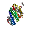
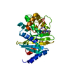
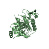
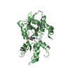
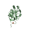
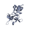
| Title | Influenza B/Memphis/13/03 endonuclease with I38T mutation | ||||||
 Components Components | Polymerase acidic protein | ||||||
 Keywords Keywords | VIRAL PROTEIN / Influenza polymerase / endonuclease | ||||||
| Function / homology |  Function and homology information Function and homology informationcap snatching / symbiont-mediated suppression of host mRNA transcription via inhibition of RNA polymerase II activity / endonuclease activity / Hydrolases; Acting on ester bonds / host cell cytoplasm / symbiont-mediated suppression of host gene expression / viral translational frameshifting / viral RNA genome replication / hydrolase activity / DNA-templated transcription ...cap snatching / symbiont-mediated suppression of host mRNA transcription via inhibition of RNA polymerase II activity / endonuclease activity / Hydrolases; Acting on ester bonds / host cell cytoplasm / symbiont-mediated suppression of host gene expression / viral translational frameshifting / viral RNA genome replication / hydrolase activity / DNA-templated transcription / host cell nucleus / RNA binding / metal ion binding Similarity search - Function | ||||||
| Biological species |  Influenza B virus Influenza B virus | ||||||
| Method |  X-RAY DIFFRACTION / SYNCHROTRON / MOLECULAR REPLACEMENT / Resolution: 1.8 Å X-RAY DIFFRACTION / SYNCHROTRON / MOLECULAR REPLACEMENT / Resolution: 1.8 Å | ||||||
 Authors Authors | Cusack, S. / Speranzini, V. | ||||||
 Citation Citation | Journal: Sci Rep / Year: 2018 Title: Characterization of influenza virus variants induced by treatment with the endonuclease inhibitor baloxavir marboxil. Authors: Omoto, S. / Speranzini, V. / Hashimoto, T. / Noshi, T. / Yamaguchi, H. / Kawai, M. / Kawaguchi, K. / Uehara, T. / Shishido, T. / Naito, A. / Cusack, S. | ||||||
| History |
|
- Structure visualization
| Structure viewer | Molecule: MolmilJmol/JSmol |
|---|
- Downloads & links
-Download
| PDBx/mmCIF format | 6fsb.cif.gz | 98.9 KB | Display | PDBx/mmCIF format |
|---|---|---|---|---|
| PDB format | pdb6fsb.ent.gz | 75 KB | Display | PDB format |
| PDBx/mmJSON format | 6fsb.json.gz | Tree view | PDBx/mmJSON format | |
| Others |  Other downloads Other downloads |
-Validation report
| Arichive directory | https://data.pdbj.org/pub/pdb/validation_reports/fs/6fsbftp://data.pdbj.org/pub/pdb/validation_reports/fs/6fsb | HTTPS FTP |
|---|
-Related structure data
| Related structure data |  6fs6C  6fs7C  6fs8C  6fs9C  5fmlS S: Starting model for refinement C: citing same article ( |
|---|---|
| Similar structure data |


-Links
 PDBj
PDBj- Assembly
| Deposited unit | 
| ||||||||
|---|---|---|---|---|---|---|---|---|---|
| 1 | 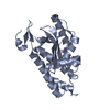
| ||||||||
| 2 | 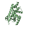
| ||||||||
| Unit cell |
|
-Components
| #1: Protein | Mass: 23506.596 Da / Num. of mol.: 2 / Mutation: I38T Source method: isolated from a genetically manipulated source Details: N-terminal GAMGSGMA linkerI38T mutation / Source: (gene. exp.) Influenza B virus (B/Memphis/13/2003) / Gene: PA / Plasmid: pETM11 / Production host:  References: UniProt: Q5V8Z9, Hydrolases; Acting on ester bonds #2: Chemical |   Mass: 54.938 Da / Num. of mol.: 2 / Source method: obtained synthetically / Formula: Mn Mass: 54.938 Da / Num. of mol.: 2 / Source method: obtained synthetically / Formula: Mn#3: Chemical | ChemComp-MG / |   Mass: 24.305 Da / Num. of mol.: 1 / Source method: obtained synthetically / Formula: Mg Mass: 24.305 Da / Num. of mol.: 1 / Source method: obtained synthetically / Formula: Mg#4: Water | ChemComp-HOH / |  Mass: 18.015 Da / Num. of mol.: 125 / Source method: isolated from a natural source / Formula: H2O Mass: 18.015 Da / Num. of mol.: 125 / Source method: isolated from a natural source / Formula: H2O |
|---|
-Experimental details
-Experiment
| Experiment | Method: X-RAY DIFFRACTION / Number of used crystals: 1 |
|---|
- Sample preparation
| Crystal | Density Matthews: 2.29 Å3/Da / Density % sol: 46.26 % |
|---|---|
| Crystal grow | Temperature: 293 K / Method: vapor diffusion, sitting drop Details: Protein, at 15-17 mg/ml, was incubated with 10-fold molar excess of BXA for 30 min at RT, mixtures were centrifuged at RT for 5 minutes at 12000 g, and soluble fraction was used for ...Details: Protein, at 15-17 mg/ml, was incubated with 10-fold molar excess of BXA for 30 min at RT, mixtures were centrifuged at RT for 5 minutes at 12000 g, and soluble fraction was used for crystallization trials (final protein concentration 8-10 mg/ml). Mother liquor was |
-Data collection
| Diffraction | Mean temperature: 100 K |
|---|---|
| Diffraction source | Source: SYNCHROTRON / Site: ESRF  / Beamline: MASSIF-1 / Wavelength: 0.966 Å / Beamline: MASSIF-1 / Wavelength: 0.966 Å |
| Detector | Type: DECTRIS PILATUS3 2M / Detector: PIXEL / Date: Oct 4, 2017 |
| Radiation | Protocol: SINGLE WAVELENGTH / Monochromatic (M) / Laue (L): M / Scattering type: x-ray |
| Radiation wavelength | Wavelength: 0.966 Å / Relative weight: 1 |
| Reflection | Resolution: 1.8→50 Å / Num. obs: 39336 / % possible obs: 91.3 % / Redundancy: 1.7 % / CC1/2: 0.996 / Rsym value: 0.055 / Net I/σ(I): 7.55 |
| Reflection shell | Resolution: 1.8→1.85 Å / Redundancy: 1.7 % / Mean I/σ(I) obs: 1.17 / Num. unique obs: 5269 / CC1/2: 0.596 / Rsym value: 0.58 / % possible all: 92.4 |
- Processing
| Software |
| ||||||||||||||||||||||||||||||||||||||||||||||||||||||||||||||||||||||||||||||||||||||||||||||||||
|---|---|---|---|---|---|---|---|---|---|---|---|---|---|---|---|---|---|---|---|---|---|---|---|---|---|---|---|---|---|---|---|---|---|---|---|---|---|---|---|---|---|---|---|---|---|---|---|---|---|---|---|---|---|---|---|---|---|---|---|---|---|---|---|---|---|---|---|---|---|---|---|---|---|---|---|---|---|---|---|---|---|---|---|---|---|---|---|---|---|---|---|---|---|---|---|---|---|---|---|
| Refinement | Method to determine structure: MOLECULAR REPLACEMENT Starting model: 5FML Resolution: 1.8→46.104 Å / SU ML: 0.27 / Cross valid method: FREE R-VALUE / σ(F): 1.37 / Phase error: 26.69
| ||||||||||||||||||||||||||||||||||||||||||||||||||||||||||||||||||||||||||||||||||||||||||||||||||
| Solvent computation | Shrinkage radii: 0.9 Å / VDW probe radii: 1.11 Å | ||||||||||||||||||||||||||||||||||||||||||||||||||||||||||||||||||||||||||||||||||||||||||||||||||
| Refinement step | Cycle: LAST / Resolution: 1.8→46.104 Å
| ||||||||||||||||||||||||||||||||||||||||||||||||||||||||||||||||||||||||||||||||||||||||||||||||||
| Refine LS restraints |
| ||||||||||||||||||||||||||||||||||||||||||||||||||||||||||||||||||||||||||||||||||||||||||||||||||
| LS refinement shell |
|
