 Movie
Movie Controller


+ Open data

- Basic information
| Entry | Database: PDB / ID: 1rxw | ||||||
|---|---|---|---|---|---|---|---|
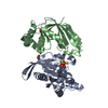
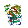
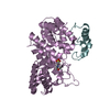
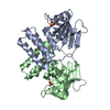
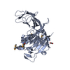
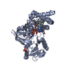
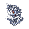
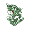
| Title | Crystal structure of A. fulgidus FEN-1 bound to DNA | ||||||
 Components Components |
| ||||||
 Keywords Keywords | HYDROLASE/DNA / helical clamp / helix-3 turn-helix / hydrophobic wedge / 3' flap binding site / HYDROLASE-DNA COMPLEX | ||||||
| Function / homology |  Function and homology information Function and homology information5'-flap endonuclease activity / DNA replication, removal of RNA primer / 5'-3' exonuclease activity / Hydrolases; Acting on ester bonds / DNA repair / magnesium ion binding / DNA binding Similarity search - Function | ||||||
| Biological species |   Archaeoglobus fulgidus (archaea) Archaeoglobus fulgidus (archaea) | ||||||
| Method |  X-RAY DIFFRACTION / SYNCHROTRON / MOLECULAR REPLACEMENT / Resolution: 2 Å X-RAY DIFFRACTION / SYNCHROTRON / MOLECULAR REPLACEMENT / Resolution: 2 Å | ||||||
 Authors Authors | Chapados, B.R. / Hosfield, D.J. / Han, S. / Qiu, J. / Yelent, B. / Shen, B. / Tainer, J.A. | ||||||
 Citation Citation | Journal: Cell(Cambridge,Mass.) / Year: 2004 Title: Structural Basis for FEN-1 Substrate Specificity and PCNA-Mediated Activation in DNA Replication and Repair Authors: Chapados, B.R. / Hosfield, D.J. / Han, S. / Qiu, J. / Yelent, B. / Shen, B. / Tainer, J.A. | ||||||
| History |
|
- Structure visualization
| Structure viewer | Molecule: MolmilJmol/JSmol |
|---|
- Downloads & links
-Download
| PDBx/mmCIF format | 1rxw.cif.gz | 87.3 KB | Display | PDBx/mmCIF format |
|---|---|---|---|---|
| PDB format | pdb1rxw.ent.gz | 62.3 KB | Display | PDB format |
| PDBx/mmJSON format | 1rxw.json.gz | Tree view | PDBx/mmJSON format | |
| Others |  Other downloads Other downloads |
-Validation report
| Arichive directory | https://data.pdbj.org/pub/pdb/validation_reports/rx/1rxwftp://data.pdbj.org/pub/pdb/validation_reports/rx/1rxw | HTTPS FTP |
|---|
-Related structure data
| Related structure data |  1rwzC  1rxmC  1rxvSC  1rxzC S: Starting model for refinement C: citing same article ( |
|---|---|
| Similar structure data |


-Links
 PDBj
PDBj

- Assembly
| Deposited unit | 
| ||||||||
|---|---|---|---|---|---|---|---|---|---|
| 1 |
| ||||||||
| Unit cell |
| ||||||||
| Components on special symmetry positions |
| ||||||||
| Details | The biological assembly is a monomer in the asymmetric unit. |
-Components
| #1: DNA chain | Mass: 2426.617 Da / Num. of mol.: 1 / Source method: obtained synthetically / Details: synthetic upstream primer strand |
|---|---|
| #2: DNA chain | Mass: 2755.823 Da / Num. of mol.: 1 / Source method: obtained synthetically / Details: synthetic upstream primer strand |
| #3: Protein | Mass: 38117.398 Da / Num. of mol.: 1 Source method: isolated from a genetically manipulated source Source: (gene. exp.) Archaeoglobus fulgidus (archaea) / Gene: FEN, AF0264 / Plasmid: pET11a / Species (production host): Escherichia coli / Production host:  |
| #4: Water | ChemComp-HOH /  Mass: 18.015 Da / Num. of mol.: 152 / Source method: isolated from a natural source / Formula: H2O Mass: 18.015 Da / Num. of mol.: 152 / Source method: isolated from a natural source / Formula: H2O |
-Experimental details
-Experiment
| Experiment | Method: X-RAY DIFFRACTION / Number of used crystals: 1 |
|---|
- Sample preparation
| Crystal | Density Matthews: 2.38 Å3/Da / Density % sol: 48.36 % | |||||||||||||||||||||||||
|---|---|---|---|---|---|---|---|---|---|---|---|---|---|---|---|---|---|---|---|---|---|---|---|---|---|---|
| Crystal grow | Temperature: 293 K / Method: vapor diffusion, hanging drop / pH: 8 Details: PEG 4000, ammonium sulfate, glycerol, pH 8, VAPOR DIFFUSION, HANGING DROP, temperature 293K | |||||||||||||||||||||||||
| Components of the solutions |
| |||||||||||||||||||||||||
| Crystal grow | *PLUS Method: vapor diffusion | |||||||||||||||||||||||||
| Components of the solutions | *PLUS
|
-Data collection
| Diffraction | Mean temperature: 100 K |
|---|---|
| Diffraction source | Source: SYNCHROTRON / Site: APS  / Beamline: 14-BM-C / Wavelength: 1 Å / Beamline: 14-BM-C / Wavelength: 1 Å |
| Detector | Type: ADSC QUANTUM 4 / Detector: CCD / Date: Sep 24, 1999 |
| Radiation | Monochromator: Ge(111). conical Si/Rh mirror / Protocol: SINGLE WAVELENGTH / Monochromatic (M) / Laue (L): M / Scattering type: x-ray |
| Radiation wavelength | Wavelength: 1 Å / Relative weight: 1 |
| Reflection | Resolution: 1.9→40 Å / Num. all: 33233 / Num. obs: 33233 / % possible obs: 98.7 % / Observed criterion σ(F): 0 / Observed criterion σ(I): 0 / Rsym value: 0.056 |
| Reflection shell | Resolution: 1.9→1.97 Å / Rsym value: 0.322 / % possible all: 97.3 |
- Processing
| Software |
| |||||||||||||||||||||||||
|---|---|---|---|---|---|---|---|---|---|---|---|---|---|---|---|---|---|---|---|---|---|---|---|---|---|---|
| Refinement | Method to determine structure: MOLECULAR REPLACEMENT Starting model: PDB ENTRY 1RXV Resolution: 2→40 Å / Cross valid method: THROUGHOUT / σ(F): 2 / Stereochemistry target values: Engh & Huber
| |||||||||||||||||||||||||
| Refinement step | Cycle: LAST / Resolution: 2→40 Å
| |||||||||||||||||||||||||
| Refine LS restraints |
|
