- PDB-1rxv: Crystal Structure of A. Fulgidus FEN-1 bound to DNA -
+
Open data
ID or keywords:
Loading...
-
Basic information
Entry
Database: PDB / ID: 1rxv
Title
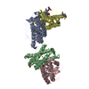
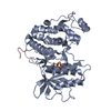
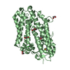
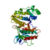
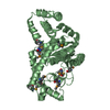
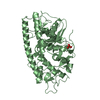
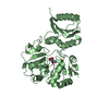
Crystal Structure of A. Fulgidus FEN-1 bound to DNA
Components
5'-d(*T*pA*pG*pC*pA*pT*pC*pG*pG)
Flap structure-specific endonuclease
Keywords
HYDROLASE/DNA / helical clamp / helix-3 turn-helix / 3' flap binding site / protein-dna complex / dna repair / dna replication / HYDROLASE-DNA COMPLEX
Function / homology
Function and homology information
5'-flap endonuclease activity / DNA replication, removal of RNA primer / 5'-3' exonuclease activity / Hydrolases; Acting on ester bonds / DNA repair / magnesium ion binding / DNA binding Similarity search - Function
SEQUENCE Authors also crystallized the sequence CGATGCTA, however no electron density was seen for ...SEQUENCE Authors also crystallized the sequence CGATGCTA, however no electron density was seen for any of the residues.
Resolution: 2.5→2.59 Å / Rsym value: 0.192 / % possible all: 97.2
-
Processing
Software
Name
Version
Classification
DENZO
datareduction
SCALEPACK
datascaling
AMoRE
phasing
CNS
1
refinement
Refinement
Method to determine structure: MOLECULAR REPLACEMENT Starting model: Model built from 2.5A resolution experimental map. Phases for this map were calculated from a four-wavelength multiple anomalous dispersion experiment. Resolution: 2.5→20 Å / Cross valid method: THROUGHOUT / σ(F): 2 / Stereochemistry target values: Engh & Huber
Rfactor
Num. reflection
Selection details
Rfree
0.309
1593
random
Rwork
0.274
-
-
obs
0.276
32212
-
all
-
33078
-
Refinement step
Cycle: LAST / Resolution: 2.5→20 Å
Protein
Nucleic acid
Ligand
Solvent
Total
Num. atoms
4886
82
0
0
4968
Refine LS restraints
Refine-ID
Type
Dev ideal
X-RAY DIFFRACTION
x_bond_d
0.007
X-RAY DIFFRACTION
x_angle_d
1.29
X-RAY DIFFRACTION
x_torsion_deg
20.9
X-RAY DIFFRACTION
x_torsion_impr_deg
0.91
Refinement
*PLUS
Rfactor Rfree: 0.238 / Rfactor Rwork: 0.242
Solvent computation
*PLUS
Displacement parameters
*PLUS
Refine LS restraints
*PLUS
Refine-ID
Type
Dev ideal
X-RAY DIFFRACTION
x_angle_d
X-RAY DIFFRACTION
x_angle_deg
1.29
+
About Yorodumi
-
News
-
Feb 9, 2022. New format data for meta-information of EMDB entries
New format data for meta-information of EMDB entries
Version 3 of the EMDB header file is now the official format.
The previous official version 1.9 will be removed from the archive.
In the structure databanks used in Yorodumi, some data are registered as the other names, "COVID-19 virus" and "2019-nCoV". Here are the details of the virus and the list of structure data.
Jan 31, 2019. EMDB accession codes are about to change! (news from PDBe EMDB page)
EMDB accession codes are about to change! (news from PDBe EMDB page)
The allocation of 4 digits for EMDB accession codes will soon come to an end. Whilst these codes will remain in use, new EMDB accession codes will include an additional digit and will expand incrementally as the available range of codes is exhausted. The current 4-digit format prefixed with “EMD-” (i.e. EMD-XXXX) will advance to a 5-digit format (i.e. EMD-XXXXX), and so on. It is currently estimated that the 4-digit codes will be depleted around Spring 2019, at which point the 5-digit format will come into force.
The EM Navigator/Yorodumi systems omit the EMD- prefix.
Related info.:Q: What is EMD? / ID/Accession-code notation in Yorodumi/EM Navigator
Yorodumi is a browser for structure data from EMDB, PDB, SASBDB, etc.
This page is also the successor to EM Navigator detail page, and also detail information page/front-end page for Omokage search.
The word "yorodu" (or yorozu) is an old Japanese word meaning "ten thousand". "mi" (miru) is to see.
Related info.:EMDB / PDB / SASBDB / Comparison of 3 databanks / Yorodumi Search / Aug 31, 2016. New EM Navigator & Yorodumi / Yorodumi Papers / Jmol/JSmol / Function and homology information / Changes in new EM Navigator and Yorodumi
 Movie
Movie Controller


 Open data
 Basic information
 Components
 Keywords
 Function and homology information
 Archaeoglobus fulgidus (archaea)
 X-RAY DIFFRACTION /
  Authors
 Citation
 Structure visualization
 Downloads & links
 Other downloads


 PDBj
PDBj

 Assembly
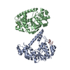


 Sample preparation
 / Beamline: 14-BM-C / Wavelength: 1 Å
 Processing