 Movie
Movie Controller


+ Open data

- Basic information
| Entry | Database: PDB / ID: 1reu | ||||||
|---|---|---|---|---|---|---|---|
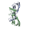
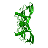
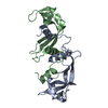
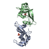
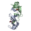
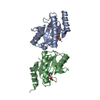
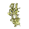
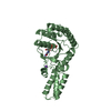
| Title | Structure of the bone morphogenetic protein 2 mutant L51P | ||||||
 Components Components | bone morphogenetic protein 2 | ||||||
 Keywords Keywords | HORMONE/GROWTH FACTOR / TGF-beta fold / HORMONE-GROWTH FACTOR COMPLEX | ||||||
| Function / homology |  Function and homology information Function and homology informationcardiac atrium formation / cardiocyte differentiation / negative regulation of calcium-independent cell-cell adhesion / positive regulation of phosphatase activity / cardiac jelly development / negative regulation of aldosterone biosynthetic process / negative regulation of cortisol biosynthetic process / positive regulation of extracellular matrix constituent secretion / atrioventricular canal morphogenesis / negative regulation of steroid biosynthetic process ...cardiac atrium formation / cardiocyte differentiation / negative regulation of calcium-independent cell-cell adhesion / positive regulation of phosphatase activity / cardiac jelly development / negative regulation of aldosterone biosynthetic process / negative regulation of cortisol biosynthetic process / positive regulation of extracellular matrix constituent secretion / atrioventricular canal morphogenesis / negative regulation of steroid biosynthetic process / embryonic heart tube anterior/posterior pattern specification / mesenchymal cell proliferation involved in ureteric bud development / regulation of odontogenesis of dentin-containing tooth / corticotropin hormone secreting cell differentiation / negative regulation of cardiac muscle cell differentiation / thyroid-stimulating hormone-secreting cell differentiation / endodermal-mesodermal cell signaling / mesenchyme development / negative regulation of insulin-like growth factor receptor signaling pathway / ameloblast differentiation / pericardium development / telencephalon regionalization / aortic valve development / positive regulation of odontogenesis / heart induction / positive regulation of cartilage development / positive regulation of peroxisome proliferator activated receptor signaling pathway / BMP receptor complex / co-receptor binding / cardiac epithelial to mesenchymal transition / lung vasculature development / mesenchymal cell differentiation / proteoglycan metabolic process / positive regulation of bone mineralization involved in bone maturation / positive regulation of odontoblast differentiation / Transcriptional regulation by RUNX2 / positive regulation of astrocyte differentiation / phosphatase activator activity / BMP receptor binding / endocardial cushion formation / telencephalon development / cellular response to BMP stimulus / Signaling by BMP / astrocyte differentiation / cardiac muscle tissue morphogenesis / cardiac muscle cell differentiation / positive regulation of ossification / positive regulation of p38MAPK cascade / atrioventricular valve morphogenesis / endocardial cushion morphogenesis / Molecules associated with elastic fibres / branching involved in ureteric bud morphogenesis / positive regulation of osteoblast proliferation / negative regulation of fat cell differentiation / odontogenesis of dentin-containing tooth / bone mineralization / inner ear development / ciliary transition zone / negative regulation of cell cycle / positive regulation of SMAD protein signal transduction / epithelial to mesenchymal transition / chondrocyte differentiation / cell fate commitment / positive regulation of osteoblast differentiation / positive regulation of bone mineralization / BMP signaling pathway / positive regulation of epithelial to mesenchymal transition / positive regulation of fat cell differentiation / positive regulation of Wnt signaling pathway / heart morphogenesis / Notch signaling pathway / positive regulation of neuron differentiation / osteoclast differentiation / cytokine activity / animal organ morphogenesis / protein serine/threonine kinase activator activity / growth factor activity / response to bacterium / skeletal system development / negative regulation of smooth muscle cell proliferation / negative regulation of transforming growth factor beta receptor signaling pathway / negative regulation of canonical Wnt signaling pathway / positive regulation of protein phosphorylation / bone development / positive regulation of miRNA transcription / protein destabilization / protein import into nucleus / osteoblast differentiation / Regulation of RUNX2 expression and activity / MAPK cascade / cell-cell signaling / heart development / transcription by RNA polymerase II / in utero embryonic development / response to hypoxia / positive regulation of ERK1 and ERK2 cascade / positive regulation of MAPK cascade / cilium / positive regulation of cell migration / positive regulation of apoptotic process Similarity search - Function | ||||||
| Biological species |  Homo sapiens (human) Homo sapiens (human) | ||||||
| Method |  X-RAY DIFFRACTION / MOLECULAR REPLACEMENT / Resolution: 2.65 Å X-RAY DIFFRACTION / MOLECULAR REPLACEMENT / Resolution: 2.65 Å | ||||||
 Authors Authors | Keller, S. / Nickel, J. / Zhang, J.-L. / Sebald, W. / Mueller, T.D. | ||||||
 Citation Citation | Journal: Nat.Struct.Mol.Biol. / Year: 2004 Title: Molecular recognition of BMP-2 and BMP receptor IA. Authors: Keller, S. / Nickel, J. / Zhang, J.L. / Sebald, W. / Mueller, T.D. #1: Journal: J.Mol.Biol. / Year: 1999Title: Crystal structure of human bone morphogenetic protein-2 at 2.7 A resolution Authors: Scheufler, C. / Sebald, W. / Huelsmeyer, M. #2: Journal: Nat.Struct.Biol. / Year: 2000Title: Crystal structure of the BMP-2-BRIA ectodomain complex Authors: Kirsch, T. / Sebald, W. / Dreyer, M.K. | ||||||
| History |
|
- Structure visualization
| Structure viewer | Molecule: MolmilJmol/JSmol |
|---|
- Downloads & links
-Download
| PDBx/mmCIF format | 1reu.cif.gz | 34.2 KB | Display | PDBx/mmCIF format |
|---|---|---|---|---|
| PDB format | pdb1reu.ent.gz | 22.7 KB | Display | PDB format |
| PDBx/mmJSON format | 1reu.json.gz | Tree view | PDBx/mmJSON format | |
| Others |  Other downloads Other downloads |
-Validation report
| Arichive directory | https://data.pdbj.org/pub/pdb/validation_reports/re/1reuftp://data.pdbj.org/pub/pdb/validation_reports/re/1reu | HTTPS FTP |
|---|
-Related structure data
| Related structure data |  1rewC  3bmpS S: Starting model for refinement C: citing same article ( |
|---|---|
| Similar structure data |


-Links
 PDBj
PDBj


- Assembly
| Deposited unit | 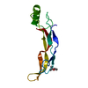
| ||||||||||
|---|---|---|---|---|---|---|---|---|---|---|---|
| 1 | 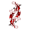
| ||||||||||
| 2 | x 6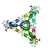
| ||||||||||
| Unit cell |
| ||||||||||
| Details | The biological assembly is a dimer generated by the two fold axis (disulfide bonded homodimer): 2/3+x-y, 1/3-y, 1/3-z |
-Components
| #1: Protein | Mass: 11498.064 Da / Num. of mol.: 1 / Fragment: mature part / Mutation: L51P Source method: isolated from a genetically manipulated source Source: (gene. exp.) Homo sapiens (human) / Plasmid: pN25c109 / Production host:  | ||||
|---|---|---|---|---|---|
| #2: Chemical |   Mass: 118.174 Da / Num. of mol.: 2 / Source method: obtained synthetically / Formula: C6H14O2 / Comment: precipitant*YM Mass: 118.174 Da / Num. of mol.: 2 / Source method: obtained synthetically / Formula: C6H14O2 / Comment: precipitant*YM#3: Water | ChemComp-HOH / |  Mass: 18.015 Da / Num. of mol.: 13 / Source method: isolated from a natural source / Formula: H2O Mass: 18.015 Da / Num. of mol.: 13 / Source method: isolated from a natural source / Formula: H2OHas protein modification | Y | |
-Experimental details
-Experiment
| Experiment | Method: X-RAY DIFFRACTION / Number of used crystals: 2 |
|---|
- Sample preparation
| Crystal | Density Matthews: 3.77 Å3/Da / Density % sol: 67.1 % |
|---|---|
| Crystal grow | Temperature: 298 K / Method: vapor diffusion, hanging drop / pH: 5 Details: lithium sulfate, tert-butanol, pH 5.0, VAPOR DIFFUSION, HANGING DROP, temperature 298K |
-Data collection
| Diffraction | Mean temperature: 298 K |
|---|---|
| Diffraction source | Source: ROTATING ANODE / Type: RIGAKU RU300 / Wavelength: 1.5418 Å |
| Detector | Type: RIGAKU RAXIS IV / Detector: IMAGE PLATE / Date: Apr 10, 2003 / Details: Osmic ConfocalBlue |
| Radiation | Monochromator: Mirrors / Protocol: SINGLE WAVELENGTH / Monochromatic (M) / Laue (L): M / Scattering type: x-ray |
| Radiation wavelength | Wavelength: 1.5418 Å / Relative weight: 1 |
| Reflection | Resolution: 2.65→19.76 Å / Num. all: 5624 / Num. obs: 5624 / % possible obs: 93.5 % / Observed criterion σ(I): 1.2 / Redundancy: 3.4 % / Biso Wilson estimate: 101.2 Å2 / Rmerge(I) obs: 0.06 |
| Reflection shell | Resolution: 2.65→2.82 Å / Mean I/σ(I) obs: 2.4 / Rsym value: 0.293 / % possible all: 72.3 |
- Processing
| Software |
| ||||||||||||||||||||||||||||||||||||
|---|---|---|---|---|---|---|---|---|---|---|---|---|---|---|---|---|---|---|---|---|---|---|---|---|---|---|---|---|---|---|---|---|---|---|---|---|---|
| Refinement | Method to determine structure: MOLECULAR REPLACEMENT Starting model: PDB ENTRY 3BMP Resolution: 2.65→19.76 Å / Rfactor Rfree error: 0.015 / Data cutoff high absF: 1489344.52 / Data cutoff low absF: 0 / Isotropic thermal model: Isotropic / Cross valid method: THROUGHOUT / σ(F): 0 / Stereochemistry target values: Engh & Huber
| ||||||||||||||||||||||||||||||||||||
| Solvent computation | Solvent model: FLAT MODEL / Bsol: 48.4107 Å2 / ksol: 0.310865 e/Å3 | ||||||||||||||||||||||||||||||||||||
| Displacement parameters | Biso mean: 64.6 Å2
| ||||||||||||||||||||||||||||||||||||
| Refine analyze |
| ||||||||||||||||||||||||||||||||||||
| Refinement step | Cycle: LAST / Resolution: 2.65→19.76 Å
| ||||||||||||||||||||||||||||||||||||
| Refine LS restraints |
| ||||||||||||||||||||||||||||||||||||
| LS refinement shell | Resolution: 2.65→2.82 Å / Rfactor Rfree error: 0.057 / Total num. of bins used: 6
| ||||||||||||||||||||||||||||||||||||
| Xplor file |
|
