 Movie
Movie Controller


+ Open data

- Basic information
| Entry | Database: PDB / ID: 1es7 | ||||||
|---|---|---|---|---|---|---|---|
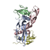
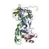
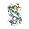
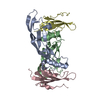
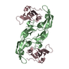
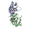
| Title | COMPLEX BETWEEN BMP-2 AND TWO BMP RECEPTOR IA ECTODOMAINS | ||||||
 Components Components |
| ||||||
 Keywords Keywords | CYTOKINE / protein-protein complex / three finger toxin fold / receptor-ligand complex / cytokine receptor / TGF beta superfamily | ||||||
| Function / homology |  Function and homology information Function and homology informationneural plate mediolateral regionalization / paraxial mesoderm structural organization / positive regulation of cardiac ventricle development / fibrous ring of heart morphogenesis / heart formation / cardiac atrium formation / positive regulation of transforming growth factor beta2 production / cardiocyte differentiation / negative regulation of calcium-independent cell-cell adhesion / positive regulation of phosphatase activity ...neural plate mediolateral regionalization / paraxial mesoderm structural organization / positive regulation of cardiac ventricle development / fibrous ring of heart morphogenesis / heart formation / cardiac atrium formation / positive regulation of transforming growth factor beta2 production / cardiocyte differentiation / negative regulation of calcium-independent cell-cell adhesion / positive regulation of phosphatase activity / cardiac jelly development / Mullerian duct regression / negative regulation of aldosterone biosynthetic process / negative regulation of cortisol biosynthetic process / atrioventricular node cell development / positive regulation of extracellular matrix constituent secretion / atrioventricular canal morphogenesis / negative regulation of steroid biosynthetic process / embryonic heart tube anterior/posterior pattern specification / mesendoderm development / mesenchymal cell proliferation involved in ureteric bud development / anti-Mullerian hormone receptor signaling pathway / regulation of odontogenesis of dentin-containing tooth / dorsal aorta morphogenesis / tricuspid valve morphogenesis / corticotropin hormone secreting cell differentiation / negative regulation of cardiac muscle cell differentiation / thyroid-stimulating hormone-secreting cell differentiation / endodermal-mesodermal cell signaling / mesenchyme development / cardiac right ventricle morphogenesis / negative regulation of insulin-like growth factor receptor signaling pathway / ameloblast differentiation / pericardium development / telencephalon regionalization / aortic valve development / hindlimb morphogenesis / BMP binding / regulation of cardiac muscle cell proliferation / negative regulation of muscle cell differentiation / atrioventricular valve development / pharyngeal arch artery morphogenesis / positive regulation of cartilage development / heart induction / positive regulation of odontogenesis / regulation of lateral mesodermal cell fate specification / positive regulation of peroxisome proliferator activated receptor signaling pathway / lateral mesoderm development / pituitary gland development / mitral valve morphogenesis / BMP receptor complex / BMP receptor activity / ventricular compact myocardium morphogenesis / negative regulation of smooth muscle cell migration / dorsal/ventral axis specification / co-receptor binding / regulation of cellular senescence / lung vasculature development / cardiac epithelial to mesenchymal transition / transforming growth factor beta receptor activity, type I / mesenchymal cell differentiation / ectoderm development / neural crest cell development / proteoglycan metabolic process / positive regulation of bone mineralization involved in bone maturation / positive regulation of odontoblast differentiation / Transcriptional regulation by RUNX2 / positive regulation of astrocyte differentiation / cardiac conduction system development / phosphatase activator activity / BMP receptor binding / endocardial cushion formation / receptor protein serine/threonine kinase / telencephalon development / cellular response to BMP stimulus / Signaling by BMP / transmembrane receptor protein serine/threonine kinase activity / ventricular trabecula myocardium morphogenesis / astrocyte differentiation / cardiac muscle tissue morphogenesis / outflow tract septum morphogenesis / dorsal/ventral pattern formation / cardiac muscle cell differentiation / positive regulation of ossification / central nervous system neuron differentiation / positive regulation of p38MAPK cascade / atrioventricular valve morphogenesis / positive regulation of dendrite development / positive regulation of mesenchymal cell proliferation / Molecules associated with elastic fibres / endocardial cushion morphogenesis / embryonic digit morphogenesis / branching involved in ureteric bud morphogenesis / ventricular septum morphogenesis / positive regulation of osteoblast proliferation / negative regulation of fat cell differentiation / odontogenesis of dentin-containing tooth / bone mineralization / SMAD binding / inner ear development Similarity search - Function | ||||||
| Biological species |  Homo sapiens (human) Homo sapiens (human) | ||||||
| Method |  X-RAY DIFFRACTION / Resolution: 2.9 Å X-RAY DIFFRACTION / Resolution: 2.9 Å | ||||||
 Authors Authors | Kirsch, T. / Sebald, W. / Dreyer, M.K. | ||||||
 Citation Citation | Journal: Nat.Struct.Biol. / Year: 2000 Title: Crystal structure of the BMP-2-BRIA ectodomain complex. Authors: Kirsch, T. / Sebald, W. / Dreyer, M.K. #1: Journal: J.Mol.Biol. / Year: 1999Title: Crystal Structure of Human Bone Morphogenetic Protein-2 at 2.7 A Resolution Authors: Scheufler, C. / Sebald, W. / Hulsmeyer, M. #2: Journal: FEBS Lett. / Year: 2000Title: Isolation of Recombinant BMP Receptor IA Ectodomain and its 2:1 Complex with BMP-2 Authors: Kirsch, T. / Nickel, J. / Sebald, W. | ||||||
| History |
|
- Structure visualization
| Structure viewer | Molecule: MolmilJmol/JSmol |
|---|
- Downloads & links
-Download
| PDBx/mmCIF format | 1es7.cif.gz | 86.4 KB | Display | PDBx/mmCIF format |
|---|---|---|---|---|
| PDB format | pdb1es7.ent.gz | 66.1 KB | Display | PDB format |
| PDBx/mmJSON format | 1es7.json.gz | Tree view | PDBx/mmJSON format | |
| Others |  Other downloads Other downloads |
-Validation report
| Arichive directory | https://data.pdbj.org/pub/pdb/validation_reports/es/1es7ftp://data.pdbj.org/pub/pdb/validation_reports/es/1es7 | HTTPS FTP |
|---|
-Related structure data
| Related structure data | |
|---|---|
| Similar structure data |


-Links
 PDBj
PDBj


- Assembly
| Deposited unit | 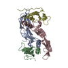
| ||||||||
|---|---|---|---|---|---|---|---|---|---|
| 1 |
| ||||||||
| Unit cell |
| ||||||||
| Details | The biological assembly is constructed from all four chains in the asymmetric unit and contains one covalently linked BMP-2 dimer and two receptor chains |
-Components
| #1: Protein | Mass: 13126.128 Da / Num. of mol.: 2 Source method: isolated from a genetically manipulated source Source: (gene. exp.) Homo sapiens (human) / Plasmid: RBSIIP / Production host:  #2: Protein | Mass: 9902.242 Da / Num. of mol.: 2 / Fragment: EXTRACELLULAR DOMAIN Source method: isolated from a genetically manipulated source Source: (gene. exp.) Homo sapiens (human) / Plasmid: PET32A / Production host: References: UniProt: P36894, Transferases; Transferring phosphorus-containing groups; Phosphotransferases with an alcohol group as acceptor #3: Water | ChemComp-HOH / |  Mass: 18.015 Da / Num. of mol.: 82 / Source method: isolated from a natural source / Formula: H2O Mass: 18.015 Da / Num. of mol.: 82 / Source method: isolated from a natural source / Formula: H2OHas protein modification | Y | |
|---|
-Experimental details
-Experiment
| Experiment | Method: X-RAY DIFFRACTION / Number of used crystals: 1 |
|---|
- Sample preparation
| Crystal | Density Matthews: 3.2 Å3/Da / Density % sol: 61 % | ||||||||||||||||||||||||||||||||||||
|---|---|---|---|---|---|---|---|---|---|---|---|---|---|---|---|---|---|---|---|---|---|---|---|---|---|---|---|---|---|---|---|---|---|---|---|---|---|
| Crystal grow | Temperature: 296 K / Method: vapor diffusion, hanging drop / pH: 7 Details: sodium acetate, imidazole, pH 7.0, VAPOR DIFFUSION, HANGING DROP, temperature 296K | ||||||||||||||||||||||||||||||||||||
| Crystal grow | *PLUS pH: 6 | ||||||||||||||||||||||||||||||||||||
| Components of the solutions | *PLUS
|
-Data collection
| Diffraction | Mean temperature: 298 K |
|---|---|
| Diffraction source | Source: ROTATING ANODE / Type: RIGAKU RU200 / Wavelength: 1.5418 |
| Detector | Type: SIEMENS X1000 / Detector: AREA DETECTOR / Date: Aug 12, 1999 |
| Radiation | Protocol: SINGLE WAVELENGTH / Monochromatic (M) / Laue (L): M / Scattering type: x-ray |
| Radiation wavelength | Wavelength: 1.5418 Å / Relative weight: 1 |
| Reflection | Resolution: 2.9→16 Å / Num. all: 52517 / Num. obs: 57236 / % possible obs: 98.6 % / Observed criterion σ(F): 0 / Observed criterion σ(I): 0 / Redundancy: 3.45 % / Rmerge(I) obs: 0.098 / Net I/σ(I): 10.2 |
| Reflection shell | Resolution: 2.9→3 Å / Redundancy: 2.9 % / Rmerge(I) obs: 0.385 / Mean I/σ(I) obs: 2.4 / Num. unique all: 1467 / % possible all: 99.5 |
| Reflection | *PLUS Num. obs: 15184 |
| Reflection shell | *PLUS % possible obs: 99.5 % |
- Processing
| Software |
| |||||||||||||||||||||||||
|---|---|---|---|---|---|---|---|---|---|---|---|---|---|---|---|---|---|---|---|---|---|---|---|---|---|---|
| Refinement | Resolution: 2.9→100 Å / σ(F): 0 / σ(I): 0 / Stereochemistry target values: Engh & Huber
| |||||||||||||||||||||||||
| Displacement parameters | Biso mean: 40 Å2 | |||||||||||||||||||||||||
| Refine analyze |
| |||||||||||||||||||||||||
| Refinement step | Cycle: LAST / Resolution: 2.9→100 Å
| |||||||||||||||||||||||||
| Refine LS restraints |
| |||||||||||||||||||||||||
| LS refinement shell | Resolution: 2.9→3 Å /
| |||||||||||||||||||||||||
| Software | *PLUS Name: CNS / Version: 0.9 / Classification: refinement | |||||||||||||||||||||||||
| Refine LS restraints | *PLUS
|
