 Movie
Movie Controller


+ Open data

- Basic information
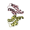
| Entry | Database: PDB / ID: 1qs7 | ||||||
|---|---|---|---|---|---|---|---|
| Title | The 1.8 angstrom structure of calmodulin rs20 peptide complex | ||||||
 Components Components |
| ||||||
 Keywords Keywords | METAL BINDING PROTEIN/PEPTIDE / CALMODULIN / CALCIUM BINDING / HELIX-LOOP-HELIX / SIGNALLING / COMPLEX(CALCIUM-BINDING PROTEIN-PEPTIDE) / METAL BINDING PROTEIN-PEPTIDE COMPLEX | ||||||
| Function / homology |  Function and homology information Function and homology informationtonic smooth muscle contraction / myosin-light-chain kinase / myosin light chain kinase activity / positive regulation of sarcomere organization / positive regulation of striated muscle contraction / myosin II complex / cardiac muscle cell development / cleavage furrow / heart morphogenesis / stress fiber ...tonic smooth muscle contraction / myosin-light-chain kinase / myosin light chain kinase activity / positive regulation of sarcomere organization / positive regulation of striated muscle contraction / myosin II complex / cardiac muscle cell development / cleavage furrow / heart morphogenesis / stress fiber / lamellipodium / calmodulin binding / calcium ion binding / ATP binding / metal ion binding / cytoplasm / cytosol Similarity search - Function | ||||||
| Biological species |  | ||||||
| Method |  X-RAY DIFFRACTION / MOLECULAR REPLACEMENT / Resolution: 1.8 Å X-RAY DIFFRACTION / MOLECULAR REPLACEMENT / Resolution: 1.8 Å | ||||||
 Authors Authors | Weigand, S. / Anderson, W.F. | ||||||
 Citation Citation | Journal: To be Published Title: High Resolution Structure of a Calmodulin Rs20 Peptide Complex Authors: Weigand, S. / Shuvalova, L. / Lukas, T.J. / Mirzoeva, S. / Watterson, D.M. / Anderson, W.F. #1: Journal: Biochemistry / Year: 1999Title: Analysis of the Functional Coupling between Calmodulin's Calcium Binding and Peptide Recognition Properties Authors: Mirzoeva, S. / Weigand, S. / Lukas, T.J. / Shuvalova, L. / Anderson, W.F. / Watterson, D.M. #2: Journal: Science / Year: 1992Title: Target Enzyme Recognition by Calmodulin: 2.4 A Structure of a Calmodulin- Peptide Complex Authors: Meador, W.E. / Means, A.R. / Quiocho, F.A. | ||||||
| History |
|
- Structure visualization
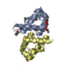
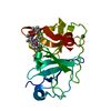
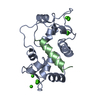
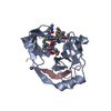
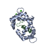
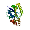
| Structure viewer | Molecule: MolmilJmol/JSmol |
|---|
- Downloads & links
-Download
| PDBx/mmCIF format | 1qs7.cif.gz | 87.3 KB | Display | PDBx/mmCIF format |
|---|---|---|---|---|
| PDB format | pdb1qs7.ent.gz | 64.8 KB | Display | PDB format |
| PDBx/mmJSON format | 1qs7.json.gz | Tree view | PDBx/mmJSON format | |
| Others |  Other downloads Other downloads |
-Validation report
| Arichive directory | https://data.pdbj.org/pub/pdb/validation_reports/qs/1qs7ftp://data.pdbj.org/pub/pdb/validation_reports/qs/1qs7 | HTTPS FTP |
|---|
-Related structure data
| Related structure data | 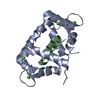 1qtxC 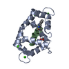 1vrkS S: Starting model for refinement C: citing same article ( |
|---|---|
| Similar structure data |


-Links
 PDBj
PDBj


- Assembly
| Deposited unit | 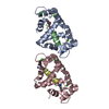
| ||||||||
|---|---|---|---|---|---|---|---|---|---|
| 1 | 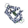
| ||||||||
| 2 | 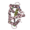
| ||||||||
| Unit cell |
|
-Components
| #1: Protein | Mass: 16327.974 Da / Num. of mol.: 2 Source method: isolated from a genetically manipulated source Source: (gene. exp.) #2: Protein/peptide | Mass: 2327.696 Da / Num. of mol.: 2 / Source method: obtained synthetically Details: PEPTIDE ANALOG OF THE CALMODULIN RECOGNITION REGION OF CHICKEN SMOOTH MUSCLE/ NONMUSCLE MYOSIN LIGHT CHAIN KINASE References: UniProt: P19038, UniProt: P11799*PLUS #3: Chemical | ChemComp-CA /   Mass: 40.078 Da / Num. of mol.: 8 / Source method: obtained synthetically / Formula: Ca Mass: 40.078 Da / Num. of mol.: 8 / Source method: obtained synthetically / Formula: Ca#4: Water | ChemComp-HOH / |  Mass: 18.015 Da / Num. of mol.: 264 / Source method: isolated from a natural source / Formula: H2O Mass: 18.015 Da / Num. of mol.: 264 / Source method: isolated from a natural source / Formula: H2OCompound details | THE PRESENCE OR ABSENCE OF THE FORMYL BLOCKING GROUP ON TRYPTOPHAN-4 OF THE PEPTIDE HAS NO EFFECT ...THE PRESENCE OR ABSENCE OF THE FORMYL BLOCKING GROUP ON TRYPTOPHAN | Has protein modification | Y | |
|---|
-Experimental details
-Experiment
| Experiment | Method: X-RAY DIFFRACTION / Number of used crystals: 1 |
|---|
- Sample preparation
| Crystal | Density Matthews: 1.94 Å3/Da / Density % sol: 36.6 % |
|---|---|
| Crystal grow | Temperature: 277 K / Method: vapor diffusion, hanging drop / pH: 4.6 Details: 20% (W/V) PEG 8000, 50 MM SODIUM ACETATE 5 MM CALCIUM CHLORIDE 0.01% (W/V) SODIUM AZIDE, pH 4.6, VAPOR DIFFUSION, HANGING DROP, temperature 277K |
-Data collection
| Diffraction | Mean temperature: 107 K |
|---|---|
| Diffraction source | Source: ROTATING ANODE / Type: RIGAKU RU200 / Wavelength: 1.5418 |
| Detector | Type: RIGAKU RAXIS IIC / Detector: IMAGE PLATE / Date: Mar 7, 1995 / Details: COLLIMATOR |
| Radiation | Monochromator: FILTER / Protocol: SINGLE WAVELENGTH / Monochromatic (M) / Laue (L): M / Scattering type: x-ray |
| Radiation wavelength | Wavelength: 1.5418 Å / Relative weight: 1 |
| Reflection | Resolution: 1.59→28.87 Å / Num. all: 39297 / Num. obs: 32557 / % possible obs: 82.8 % / Observed criterion σ(F): 0 / Observed criterion σ(I): -3 / Redundancy: 3.3 % / Biso Wilson estimate: 24 Å2 / Rmerge(I) obs: 0.042 / Net I/σ(I): 24 |
| Reflection shell | Resolution: 1.59→1.84 Å / Redundancy: 3.4 % / Rmerge(I) obs: 0.16 / Mean I/σ(I) obs: 8.6 / % possible all: 91.8 |
- Processing
| Software |
| ||||||||||||||||||||||||||||||||||||||||||||||||||||||||||||||||||||||||||||||||
|---|---|---|---|---|---|---|---|---|---|---|---|---|---|---|---|---|---|---|---|---|---|---|---|---|---|---|---|---|---|---|---|---|---|---|---|---|---|---|---|---|---|---|---|---|---|---|---|---|---|---|---|---|---|---|---|---|---|---|---|---|---|---|---|---|---|---|---|---|---|---|---|---|---|---|---|---|---|---|---|---|---|
| Refinement | Method to determine structure: MOLECULAR REPLACEMENT Starting model: PDB ENTRY 1VRK Resolution: 1.8→28.87 Å / Rfactor Rfree error: 0.005 / Data cutoff high absF: 1057648.92 / Data cutoff low absF: 0 / Isotropic thermal model: RESTRAINED / Cross valid method: THROUGHOUT / σ(F): 0 / σ(I): 0 Stereochemistry target values: ENGH, R.A. AND HUBER, R. (1991). ACTA CRYST. A47, 392-400.
| ||||||||||||||||||||||||||||||||||||||||||||||||||||||||||||||||||||||||||||||||
| Displacement parameters | Biso mean: 26 Å2
| ||||||||||||||||||||||||||||||||||||||||||||||||||||||||||||||||||||||||||||||||
| Refine analyze |
| ||||||||||||||||||||||||||||||||||||||||||||||||||||||||||||||||||||||||||||||||
| Refinement step | Cycle: LAST / Resolution: 1.8→28.87 Å
| ||||||||||||||||||||||||||||||||||||||||||||||||||||||||||||||||||||||||||||||||
| Refine LS restraints |
| ||||||||||||||||||||||||||||||||||||||||||||||||||||||||||||||||||||||||||||||||
| LS refinement shell | Resolution: 1.8→1.85 Å / Rfactor Rfree error: 0.032 / Total num. of bins used: 14
| ||||||||||||||||||||||||||||||||||||||||||||||||||||||||||||||||||||||||||||||||
| Xplor file |
|
