 Movie
Movie Controller


+ Open data

- Basic information
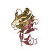
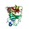
| Entry | Database: PDB / ID: 3dtj | ||||||
|---|---|---|---|---|---|---|---|
| Title | HIV-1 capsid C-terminal domain mutant (E187A) | ||||||
 Components Components | HIV-1 capsid protein | ||||||
 Keywords Keywords | VIRAL PROTEIN / HIV / CAPSID / MUTANT / INHIBITOR / ASSEMBLY / POLYPROTEIN / COMPLEX(VIRAL PROTEIN-PEPTIDE) / MAINLY ALPHA | ||||||
| Function / homology |  Function and homology information Function and homology informationviral budding via host ESCRT complex / HIV-1 retropepsin / symbiont-mediated activation of host apoptosis / retroviral ribonuclease H / exoribonuclease H / exoribonuclease H activity / DNA integration / viral genome integration into host DNA / establishment of integrated proviral latency / RNA-directed DNA polymerase ...viral budding via host ESCRT complex / HIV-1 retropepsin / symbiont-mediated activation of host apoptosis / retroviral ribonuclease H / exoribonuclease H / exoribonuclease H activity / DNA integration / viral genome integration into host DNA / establishment of integrated proviral latency / RNA-directed DNA polymerase / RNA stem-loop binding / viral penetration into host nucleus / host multivesicular body / RNA-directed DNA polymerase activity / RNA-DNA hybrid ribonuclease activity / Transferases; Transferring phosphorus-containing groups; Nucleotidyltransferases / host cell / viral nucleocapsid / DNA recombination / DNA-directed DNA polymerase / aspartic-type endopeptidase activity / Hydrolases; Acting on ester bonds / DNA-directed DNA polymerase activity / symbiont-mediated suppression of host gene expression / viral translational frameshifting / symbiont entry into host cell / lipid binding / host cell nucleus / host cell plasma membrane / virion membrane / structural molecule activity / proteolysis / DNA binding / RNA binding / zinc ion binding Similarity search - Function | ||||||
| Biological species |   Human immunodeficiency virus 1 Human immunodeficiency virus 1 | ||||||
| Method |  X-RAY DIFFRACTION / SYNCHROTRON / MOLECULAR REPLACEMENT / Resolution: 4 Å X-RAY DIFFRACTION / SYNCHROTRON / MOLECULAR REPLACEMENT / Resolution: 4 Å | ||||||
 Authors Authors | Igonet, S. / Vaney, M.C. / Rey, F.A. | ||||||
 Citation Citation | Journal: J.Biol.Chem. / Year: 2008 Title: Residues in the HIV-1 Capsid Assembly Inhibitor Binding Site Are Essential for Maintaining the Assembly-competent Quaternary Structure of the Capsid Protein. Authors: Bartonova, V. / Igonet, S. / Sticht, J. / Glass, B. / Habermann, A. / Vaney, M.C. / Sehr, P. / Lewis, J. / Rey, F.A. / Krausslich, H.G. #1: Journal: Nat.Struct.Mol.Biol. / Year: 2005Title: The HIV-1 capsid protein C-terminal domain in complex with a virus assembly inhibitor Authors: Ternois, F. / Sticht, J. / Duquerroy, S. / Krausslich, H.-G. / Rey, F.A. | ||||||
| History |
|
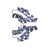
- Structure visualization
| Structure viewer | Molecule: MolmilJmol/JSmol |
|---|
- Downloads & links
-Download
| PDBx/mmCIF format | 3dtj.cif.gz | 65.9 KB | Display | PDBx/mmCIF format |
|---|---|---|---|---|
| PDB format | pdb3dtj.ent.gz | 49.8 KB | Display | PDB format |
| PDBx/mmJSON format | 3dtj.json.gz | Tree view | PDBx/mmJSON format | |
| Others |  Other downloads Other downloads |
-Validation report
| Arichive directory | https://data.pdbj.org/pub/pdb/validation_reports/dt/3dtjftp://data.pdbj.org/pub/pdb/validation_reports/dt/3dtj | HTTPS FTP |
|---|
-Related structure data
| Related structure data |  3dphC 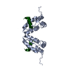 3ds0C  3ds1C 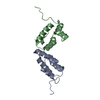 3ds2C  3ds3C  3ds4C  3ds5SC C: citing same article ( S: Starting model for refinement |
|---|---|
| Similar structure data |


-Links
 PDBj
PDBj

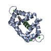
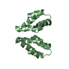
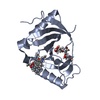
- Assembly
| Deposited unit | 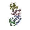
| ||||||||
|---|---|---|---|---|---|---|---|---|---|
| 1 | 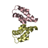
| ||||||||
| 2 | 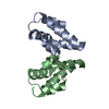
| ||||||||
| Unit cell |
|
-Components
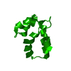
| #1: Protein | Mass: 9472.884 Da / Num. of mol.: 4 / Fragment: C-terminal domain, UNP residues 278-363 / Mutation: E187A Source method: isolated from a genetically manipulated source Source: (gene. exp.) Human immunodeficiency virus 1 / Strain: NL4-3 / Gene: gag / Plasmid: pET11c / Production host:  |
|---|
-Experimental details
-Experiment
| Experiment | Method: X-RAY DIFFRACTION / Number of used crystals: 1 |
|---|
- Sample preparation
| Crystal | Density Matthews: 2.8 Å3/Da / Density % sol: 56.14 % |
|---|---|
| Crystal grow | Temperature: 298 K / Method: evaporation / pH: 5 Details: 32% PEG 4,000, 100 mM ammonium acetate pH 5.0 and 10 mM MgCl2., EVAPORATION, temperature 298K |
-Data collection
| Diffraction | Mean temperature: 100 K |
|---|---|
| Diffraction source | Source: SYNCHROTRON / Site: SLS  / Beamline: X06SA / Wavelength: 1.044 Å / Beamline: X06SA / Wavelength: 1.044 Å |
| Detector | Type: MARMOSAIC 225 mm CCD / Detector: CCD / Date: May 25, 2005 / Details: Dynamically bendable mirror |
| Radiation | Monochromator: Si(111) monochromator / Protocol: SINGLE WAVELENGTH / Monochromatic (M) / Laue (L): M / Scattering type: x-ray |
| Radiation wavelength | Wavelength: 1.044 Å / Relative weight: 1 |
| Reflection | Resolution: 4→30.2 Å / Num. obs: 2160 / % possible obs: 62.2 % / Observed criterion σ(F): 0 / Observed criterion σ(I): 0 / Rmerge(I) obs: 0.092 / Rsym value: 0.092 / Net I/σ(I): 12.4 |
- Processing
| Software |
| ||||||||||||
|---|---|---|---|---|---|---|---|---|---|---|---|---|---|
| Refinement | Method to determine structure: MOLECULAR REPLACEMENT Starting model: 3DS5 Resolution: 4→30.2 Å / σ(F): 0 / σ(I): 0 Details: No refinement was undertaken because of the low resolution data. Only molecular replacement with the 3D model 3DS5 (HIV-1 C-terminal domain capsid mutant (N183A)) gave a solution with the ...Details: No refinement was undertaken because of the low resolution data. Only molecular replacement with the 3D model 3DS5 (HIV-1 C-terminal domain capsid mutant (N183A)) gave a solution with the same packing of the molecules. | ||||||||||||
| Refinement step | Cycle: LAST / Resolution: 4→30.2 Å
|
