 Movie
Movie Controller

[English] 日本語
 Yorodumi
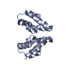
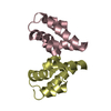
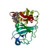
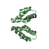
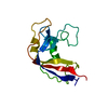
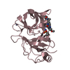
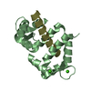
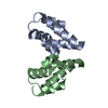
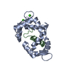
- PDB-4m0i: CRYSTAL STRUCTURE OF SYNTHETIC HIV-1 CAPSID C-TERMINAL DOMAIN (CT... -
+ Open data

- Basic information
| Entry | Database: PDB / ID: 4m0i | ||||||
|---|---|---|---|---|---|---|---|
| Title | CRYSTAL STRUCTURE OF SYNTHETIC HIV-1 CAPSID C-TERMINAL DOMAIN (CTD) C198S mutant | ||||||
 Components Components | HIV-1 CAPSID PROTEIN | ||||||
 Keywords Keywords | VIRAL PROTEIN / HIV-1 CAPSID / CORE PROTEIN / HIV-1 CAPSID C-TERMINAL DOMAIN / AIDS | ||||||
| Function / homology |  Function and homology information Function and homology informationviral nucleocapsid / host cell cytoplasm / viral translational frameshifting / host cell plasma membrane / host cell nucleus / RNA binding Similarity search - Function | ||||||
| Biological species |   Human immunodeficiency virus 1 Human immunodeficiency virus 1 | ||||||
| Method |  X-RAY DIFFRACTION / SYNCHROTRON / MOLECULAR REPLACEMENT / Resolution: 2.8 Å X-RAY DIFFRACTION / SYNCHROTRON / MOLECULAR REPLACEMENT / Resolution: 2.8 Å | ||||||
 Authors Authors | Howell, K. / Tolbert, W.D. / Pazgier, M. / Lu, W. | ||||||
 Citation Citation | Journal: To be Published Title: Molecular basis of disulfide bonding-regulated HIV-1 capsid assembly Authors: Howell, K. / Li, C. / Tolbert, W.D. / Beckett, D. / Pazgier, M. / Lu, W. | ||||||
| History |
| ||||||
| Remark 650 | HELIX DETERMINATION METHOD: AUTHOR DETERMINED |
- Structure visualization
| Structure viewer | Molecule: MolmilJmol/JSmol |
|---|
- Downloads & links
-Download
| PDBx/mmCIF format | 4m0i.cif.gz | 26.4 KB | Display | PDBx/mmCIF format |
|---|---|---|---|---|
| PDB format | pdb4m0i.ent.gz | 16.6 KB | Display | PDB format |
| PDBx/mmJSON format | 4m0i.json.gz | Tree view | PDBx/mmJSON format | |
| Others |  Other downloads Other downloads |
-Validation report
| Arichive directory | https://data.pdbj.org/pub/pdb/validation_reports/m0/4m0iftp://data.pdbj.org/pub/pdb/validation_reports/m0/4m0i | HTTPS FTP |
|---|
-Related structure data
| Related structure data |  3lryS S: Starting model for refinement |
|---|---|
| Similar structure data |

-Links
 PDBj
PDBj
- Assembly
| Deposited unit | 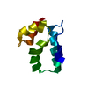
| ||||||||
|---|---|---|---|---|---|---|---|---|---|
| 1 | 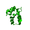
| ||||||||
| Unit cell |
|
-Components
| #1: Protein | Mass: 9514.855 Da / Num. of mol.: 1 / Fragment: C-TERMINAL DOMAIN, residues 146-231 / Mutation: C198S / Source method: obtained synthetically Details: This sequence is the HIV-1 CAPSID C-TERMINAL DOMAIN with the cysteine 198 to serine mutation Source: (synth.) Human immunodeficiency virus 1 / References: UniProt: Q71B91 |
|---|---|
| #2: Water | ChemComp-HOH /  Mass: 18.015 Da / Num. of mol.: 2 / Source method: isolated from a natural source / Formula: H2O Mass: 18.015 Da / Num. of mol.: 2 / Source method: isolated from a natural source / Formula: H2O |
-Experimental details
-Experiment
| Experiment | Method: X-RAY DIFFRACTION / Number of used crystals: 1 |
|---|
- Sample preparation
| Crystal | Density Matthews: 2.8 Å3/Da / Density % sol: 56.04 % |
|---|---|
| Crystal grow | Temperature: 294 K / Method: vapor diffusion, hanging drop / pH: 6.5 Details: 2 M ammonium sulfate, 100 mM sodium cacodylate pH 6.5, and 200 mM sodium chloride, VAPOR DIFFUSION, HANGING DROP, temperature 294K |
-Data collection
| Diffraction | Mean temperature: 100 K |
|---|---|
| Diffraction source | Source: SYNCHROTRON / Site: SSRL  / Beamline: BL7-1 / Wavelength: 0.9753 Å / Beamline: BL7-1 / Wavelength: 0.9753 Å |
| Detector | Type: MARMOSAIC 325 mm CCD / Detector: CCD / Date: May 4, 2013 / Details: RH COATED FLAT MIRROR |
| Radiation | Monochromator: SI(111) / Protocol: SINGLE WAVELENGTH / Monochromatic (M) / Laue (L): M / Scattering type: x-ray |
| Radiation wavelength | Wavelength: 0.9753 Å / Relative weight: 1 |
| Reflection | Resolution: 2.8→50 Å / Num. obs: 2648 / % possible obs: 99.1 % / Observed criterion σ(F): 0 / Observed criterion σ(I): 0 / Redundancy: 6.4 % / Rmerge(I) obs: 0.057 / Net I/σ(I): 30.7 |
| Reflection shell | Resolution: 2.8→2.85 Å / Redundancy: 6.5 % / Rmerge(I) obs: 0.669 / Mean I/σ(I) obs: 2.2 / % possible all: 99.3 |
- Processing
| Software |
| |||||||||||||||||||||||||||||||||||||||||||||||||||||||||||||||||
|---|---|---|---|---|---|---|---|---|---|---|---|---|---|---|---|---|---|---|---|---|---|---|---|---|---|---|---|---|---|---|---|---|---|---|---|---|---|---|---|---|---|---|---|---|---|---|---|---|---|---|---|---|---|---|---|---|---|---|---|---|---|---|---|---|---|---|
| Refinement | Method to determine structure: MOLECULAR REPLACEMENT Starting model: PDB ENTRY 3LRY Resolution: 2.8→30.61 Å / Cor.coef. Fo:Fc: 0.946 / Cor.coef. Fo:Fc free: 0.921 / SU B: 25.209 / SU ML: 0.42 / Cross valid method: THROUGHOUT / σ(F): 0 / ESU R: 1.086 / ESU R Free: 0.4 / Stereochemistry target values: MAXIMUM LIKELIHOOD / Details: HYDROGENS HAVE BEEN USED IF PRESENT IN THE INPUT
| |||||||||||||||||||||||||||||||||||||||||||||||||||||||||||||||||
| Solvent computation | Ion probe radii: 0.8 Å / Shrinkage radii: 0.8 Å / VDW probe radii: 1.2 Å / Solvent model: BABINET MODEL WITH MASK | |||||||||||||||||||||||||||||||||||||||||||||||||||||||||||||||||
| Displacement parameters | Biso mean: 94.951 Å2
| |||||||||||||||||||||||||||||||||||||||||||||||||||||||||||||||||
| Refinement step | Cycle: LAST / Resolution: 2.8→30.61 Å
| |||||||||||||||||||||||||||||||||||||||||||||||||||||||||||||||||
| Refine LS restraints |
| |||||||||||||||||||||||||||||||||||||||||||||||||||||||||||||||||
| LS refinement shell | Resolution: 2.8→2.872 Å / Total num. of bins used: 20
|
