 Movie
Movie Controller

[English] 日本語
 Yorodumi
- PDB-4i6u: Crystal Structure of a Y37F mutant of the Restriction-Modificatio... -
+ Open data

- Basic information
| Entry | Database: PDB / ID: 4i6u | ||||||
|---|---|---|---|---|---|---|---|
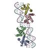
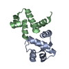
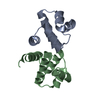
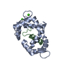
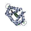
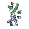
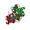
| Title | Crystal Structure of a Y37F mutant of the Restriction-Modification Controller Protein C.Esp1396I | ||||||
 Components Components | Regulatory protein | ||||||
 Keywords Keywords | TRANSCRIPTION / Restriction-modification / helix-turn-helix / transcriptional regulato / DNA | ||||||
| Function / homology |  Function and homology information Function and homology information | ||||||
| Biological species |  Enterobacter sp. (bacteria) Enterobacter sp. (bacteria) | ||||||
| Method |  X-RAY DIFFRACTION / SYNCHROTRON / MOLECULAR REPLACEMENT / Resolution: 1.97 Å X-RAY DIFFRACTION / SYNCHROTRON / MOLECULAR REPLACEMENT / Resolution: 1.97 Å | ||||||
 Authors Authors | Martin, R.N.A. / McGeehan, J.E. / Kneale, G.G. | ||||||
 Citation Citation | Journal: Plos One / Year: 2014 Title: Structural and Mutagenic Analysis of the RM Controller Protein C.Esp1396I. Authors: Martin, R.N. / McGeehan, J.E. / Kneale, G. #1: Journal: Acta Crystallogr.,Sect.D / Year: 2009Title: Structure of the restriction-modification controller protein C.Esp1396I. Authors: Ball, N. / Streeter, S.D. / Kneale, G.G. / McGeehan, J.E. #2: Journal: Nucleic Acids Res. / Year: 2012Title: Recognition of dual symmetry by the controller protein C.Esp1396I based on the structure of the transcriptional activation complex. Authors: McGeehan, J.E. / Ball, N.J. / Streeter, S.D. / Thresh, S.J. / Kneale, G.G. #3: Journal: Nucleic Acids Res. / Year: 2012Title: The structural basis of differential DNA sequence recognition by restriction-modification controller proteins. Authors: Ball, N.J. / McGeehan, J.E. / Streeter, S.D. / Thresh, S.J. / Kneale, G.G. #4: Journal: Acta Crystallogr.,Sect.D / Year: 2009Title: Structure of the restriction-modification controller protein C.Esp1396I. Authors: Ball, N. / Streeter, S.D. / Kneale, G.G. / McGeehan, J.E. | ||||||
| History |
|
- Structure visualization
| Structure viewer | Molecule: MolmilJmol/JSmol |
|---|
- Downloads & links
-Download
| PDBx/mmCIF format | 4i6u.cif.gz | 107.3 KB | Display | PDBx/mmCIF format |
|---|---|---|---|---|
| PDB format | pdb4i6u.ent.gz | 85.2 KB | Display | PDB format |
| PDBx/mmJSON format | 4i6u.json.gz | Tree view | PDBx/mmJSON format | |
| Others |  Other downloads Other downloads |
-Validation report
| Arichive directory | https://data.pdbj.org/pub/pdb/validation_reports/i6/4i6uftp://data.pdbj.org/pub/pdb/validation_reports/i6/4i6u | HTTPS FTP |
|---|
-Related structure data
| Related structure data | 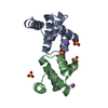 4f8dC  4fbiC 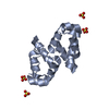 4fn3C  4i6rC 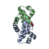 4i6tC  4ia8C  4ivzC C: citing same article ( |
|---|---|
| Similar structure data |


-Links
 PDBj
PDBj


- Assembly
| Deposited unit | 
| ||||||||
|---|---|---|---|---|---|---|---|---|---|
| 1 | 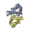
| ||||||||
| 2 | 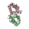
| ||||||||
| 3 | 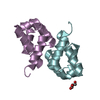
| ||||||||
| Unit cell |
|
-Components
-Protein , 1 types, 6 molecules ABCDEF
| #1: Protein | Mass: 9505.175 Da / Num. of mol.: 6 / Mutation: Y37F Source method: isolated from a genetically manipulated source Source: (gene. exp.) Enterobacter sp. (bacteria) / Strain: RFL1396 / Gene: esp1396IC / Plasmid: pET28 / Production host: |
|---|
-Non-polymers , 5 types, 155 molecules 


| #2: Chemical | ChemComp-NA /  Mass: 22.990 Da / Num. of mol.: 1 / Source method: obtained synthetically / Formula: Na Mass: 22.990 Da / Num. of mol.: 1 / Source method: obtained synthetically / Formula: Na | ||||||
|---|---|---|---|---|---|---|---|
| #3: Chemical |  Mass: 59.044 Da / Num. of mol.: 3 / Source method: obtained synthetically / Formula: C2H3O2 Mass: 59.044 Da / Num. of mol.: 3 / Source method: obtained synthetically / Formula: C2H3O2#4: Chemical |  Mass: 106.120 Da / Num. of mol.: 2 / Source method: obtained synthetically / Formula: C4H10O3 Mass: 106.120 Da / Num. of mol.: 2 / Source method: obtained synthetically / Formula: C4H10O3#5: Chemical |  Mass: 92.094 Da / Num. of mol.: 2 / Source method: obtained synthetically / Formula: C3H8O3 Mass: 92.094 Da / Num. of mol.: 2 / Source method: obtained synthetically / Formula: C3H8O3#6: Water | ChemComp-HOH / | Mass: 18.015 Da / Num. of mol.: 147 / Source method: isolated from a natural source / Formula: H2O |
-Experimental details
-Experiment
| Experiment | Method: X-RAY DIFFRACTION / Number of used crystals: 1 |
|---|
- Sample preparation
| Crystal | Density Matthews: 2.36 Å3/Da / Density % sol: 47.79 % |
|---|---|
| Crystal grow | Temperature: 277 K / Method: vapor diffusion, sitting drop / pH: 8.5 Details: 0.1 M sodium sulphate, 0.2 M sodium acetate, 0.1 M Bis Tris propane, 20 % w/v PEG 3350, pH 8.5, VAPOR DIFFUSION, SITTING DROP, temperature 277K |
-Data collection
| Diffraction | Mean temperature: 100 K |
|---|---|
| Diffraction source | Source: SYNCHROTRON / Site: Diamond  / Beamline: I04-1 / Wavelength: 0.92 Å / Beamline: I04-1 / Wavelength: 0.92 Å |
| Detector | Type: DECTRIS PILATUS 2M / Detector: PIXEL / Date: Sep 15, 2012 |
| Radiation | Monochromator: Si(111) double crystal monochromator / Protocol: SINGLE WAVELENGTH / Monochromatic (M) / Laue (L): M / Scattering type: x-ray |
| Radiation wavelength | Wavelength: 0.92 Å / Relative weight: 1 |
| Reflection | Resolution: 1.97→45.74 Å / Num. all: 527601 / Num. obs: 38881 / Rmerge(I) obs: 0.081 / Net I/σ(I): 22.6 |
- Processing
| Software |
| ||||||||||||||||||||||||||||||||||||||||||||||||||||||||||||
|---|---|---|---|---|---|---|---|---|---|---|---|---|---|---|---|---|---|---|---|---|---|---|---|---|---|---|---|---|---|---|---|---|---|---|---|---|---|---|---|---|---|---|---|---|---|---|---|---|---|---|---|---|---|---|---|---|---|---|---|---|---|
| Refinement | Method to determine structure: MOLECULAR REPLACEMENT / Resolution: 1.97→41.79 Å / Cor.coef. Fo:Fc: 0.954 / Cor.coef. Fo:Fc free: 0.928 / Occupancy max: 1 / Occupancy min: 0.5 / SU B: 3.063 / SU ML: 0.089 / Cross valid method: THROUGHOUT / σ(F): 0 / ESU R: 0.15 / ESU R Free: 0.143 / Stereochemistry target values: MAXIMUM LIKELIHOOD Details: HYDROGENS HAVE BEEN ADDED IN THE RIDING POSITIONS U VALUES : REFINED INDIVIDUALLY
| ||||||||||||||||||||||||||||||||||||||||||||||||||||||||||||
| Solvent computation | Ion probe radii: 0.8 Å / Shrinkage radii: 0.8 Å / VDW probe radii: 1.2 Å / Solvent model: MASK | ||||||||||||||||||||||||||||||||||||||||||||||||||||||||||||
| Displacement parameters | Biso max: 72.08 Å2 / Biso mean: 23.0441 Å2 / Biso min: 9.35 Å2
| ||||||||||||||||||||||||||||||||||||||||||||||||||||||||||||
| Refinement step | Cycle: LAST / Resolution: 1.97→41.79 Å
| ||||||||||||||||||||||||||||||||||||||||||||||||||||||||||||
| Refine LS restraints |
| ||||||||||||||||||||||||||||||||||||||||||||||||||||||||||||
| LS refinement shell | Resolution: 1.97→2.021 Å / Total num. of bins used: 20
|
