 Movie
Movie Controller

[English] 日本語
 Yorodumi
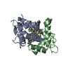
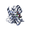
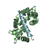
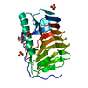
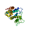
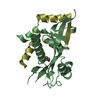
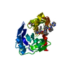
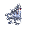
- PDB-1wrl: Crystal structure of the N-terminal domain of human cardiac tropo... -
+ Open data

- Basic information
| Entry | Database: PDB / ID: 1wrl | ||||||
|---|---|---|---|---|---|---|---|
| Title | Crystal structure of the N-terminal domain of human cardiac troponin C in complex with trifluoperazine (monoclinic crystal form) | ||||||
 Components Components | Troponin C, slow skeletal and cardiac muscles | ||||||
 Keywords Keywords | CONTRACTILE PROTEIN / EF-hand | ||||||
| Function / homology |  Function and homology information Function and homology informationdiaphragm contraction / regulation of muscle filament sliding speed / troponin T binding / cardiac Troponin complex / troponin complex / regulation of muscle contraction / transition between fast and slow fiber / Striated Muscle Contraction / muscle filament sliding / response to metal ion ...diaphragm contraction / regulation of muscle filament sliding speed / troponin T binding / cardiac Troponin complex / troponin complex / regulation of muscle contraction / transition between fast and slow fiber / Striated Muscle Contraction / muscle filament sliding / response to metal ion / cardiac muscle cell contraction / ventricular cardiac muscle tissue morphogenesis / troponin I binding / skeletal muscle contraction / cardiac muscle contraction / sarcomere / calcium-dependent protein binding / actin filament binding / calcium ion binding / protein homodimerization activity / cytosol Similarity search - Function | ||||||
| Biological species |  Homo sapiens (human) Homo sapiens (human) | ||||||
| Method |  X-RAY DIFFRACTION / SYNCHROTRON / MOLECULAR REPLACEMENT / Resolution: 2.6 Å X-RAY DIFFRACTION / SYNCHROTRON / MOLECULAR REPLACEMENT / Resolution: 2.6 Å | ||||||
 Authors Authors | Takeda, S. / Igarashi, T. / Oishi, Y. / Mori, H. | ||||||
 Citation Citation | Journal: To be Published Title: Crystal structure of the N-terminal domain of human cardiac troponin C in complex with trifluoperazine Authors: Takeda, S. / Igarashi, T. / Oishi, Y. / Mori, H. | ||||||
| History |
|
- Structure visualization
| Structure viewer | Molecule: MolmilJmol/JSmol |
|---|
- Downloads & links
-Download
| PDBx/mmCIF format | 1wrl.cif.gz | 116.5 KB | Display | PDBx/mmCIF format |
|---|---|---|---|---|
| PDB format | pdb1wrl.ent.gz | 94.1 KB | Display | PDB format |
| PDBx/mmJSON format | 1wrl.json.gz | Tree view | PDBx/mmJSON format | |
| Others |  Other downloads Other downloads |
-Validation report
| Arichive directory | https://data.pdbj.org/pub/pdb/validation_reports/wr/1wrlftp://data.pdbj.org/pub/pdb/validation_reports/wr/1wrl | HTTPS FTP |
|---|
-Related structure data

-Links
 PDBj
PDBj


- Assembly
| Deposited unit | 
| ||||||||
|---|---|---|---|---|---|---|---|---|---|
| 1 | 
| ||||||||
| 2 | 
| ||||||||
| 3 | 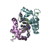
| ||||||||
| Unit cell |
|
-Components
| #1: Protein | Mass: 9951.096 Da / Num. of mol.: 6 / Fragment: N-terminal domain / Mutation: C35S, C84S Source method: isolated from a genetically manipulated source Source: (gene. exp.) Homo sapiens (human) / Plasmid: pET-3d / Production host:  #2: Chemical | ChemComp-CA /   Mass: 40.078 Da / Num. of mol.: 6 / Source method: obtained synthetically / Formula: Ca Mass: 40.078 Da / Num. of mol.: 6 / Source method: obtained synthetically / Formula: Ca#3: Chemical | ChemComp-TFP / 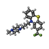  Mass: 407.496 Da / Num. of mol.: 12 / Source method: obtained synthetically / Formula: C21H24F3N3S Mass: 407.496 Da / Num. of mol.: 12 / Source method: obtained synthetically / Formula: C21H24F3N3S#4: Water | ChemComp-HOH / |  Mass: 18.015 Da / Num. of mol.: 83 / Source method: isolated from a natural source / Formula: H2O Mass: 18.015 Da / Num. of mol.: 83 / Source method: isolated from a natural source / Formula: H2O |
|---|
-Experimental details
-Experiment
| Experiment | Method: X-RAY DIFFRACTION / Number of used crystals: 1 |
|---|
- Sample preparation
| Crystal | Density Matthews: 2.5 Å3/Da / Density % sol: 49.7 % |
|---|---|
| Crystal grow | Temperature: 277 K / Method: vapor diffusion, sitting drop / pH: 7.2 Details: PEG3350, magnesium chloride, calcium chloride, MES, pH 7.2, VAPOR DIFFUSION, SITTING DROP, temperature 277K |
-Data collection
| Diffraction | Mean temperature: 90 K |
|---|---|
| Diffraction source | Source: SYNCHROTRON / Site: SPring-8  / Beamline: BL45PX / Wavelength: 1 Å / Beamline: BL45PX / Wavelength: 1 Å |
| Detector | Type: RIGAKU RAXIS V / Detector: IMAGE PLATE / Date: Feb 7, 2004 |
| Radiation | Monochromator: diamond / Protocol: SINGLE WAVELENGTH / Monochromatic (M) / Laue (L): M / Scattering type: x-ray |
| Radiation wavelength | Wavelength: 1 Å / Relative weight: 1 |
| Reflection | Resolution: 2.6→50 Å / Num. all: 17934 / Num. obs: 17906 / % possible obs: 99.8 % / Observed criterion σ(F): 0 / Observed criterion σ(I): 0 / Redundancy: 3.8 % / Rmerge(I) obs: 0.079 / Net I/σ(I): 11.6 |
| Reflection shell | Resolution: 2.6→2.69 Å / Rmerge(I) obs: 0.324 / Num. unique all: 1778 / % possible all: 99.2 |
- Processing
| Software |
| ||||||||||||||||||||
|---|---|---|---|---|---|---|---|---|---|---|---|---|---|---|---|---|---|---|---|---|---|
| Refinement | Method to determine structure: MOLECULAR REPLACEMENT / Resolution: 2.6→50 Å / Cross valid method: THROUGHOUT / σ(F): 0 / Stereochemistry target values: Engh & Huber
| ||||||||||||||||||||
| Displacement parameters | Biso mean: 51.3 Å2 | ||||||||||||||||||||
| Refinement step | Cycle: LAST / Resolution: 2.6→50 Å
| ||||||||||||||||||||
| Refine LS restraints |
| ||||||||||||||||||||
| LS refinement shell | Resolution: 2.6→2.69 Å
|
