 Movie
Movie Controller

[English] 日本語
 Yorodumi
- PDB-1w3c: Crystal structure of the Hepatitis C Virus NS3 Protease in comple... -
+ Open data

- Basic information
| Entry | Database: PDB / ID: 1w3c | ||||||
|---|---|---|---|---|---|---|---|
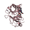
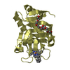
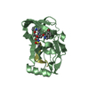
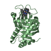
| Title | Crystal structure of the Hepatitis C Virus NS3 Protease in complex with a peptidomimetic inhibitor | ||||||
 Components Components |
| ||||||
 Keywords Keywords | HYDROLASE / SERINE PROTEASE / HCV / INDOLINE-BASED PEPTIDOMIMETIC INHIBITOR | ||||||
| Function / homology |  Function and homology information Function and homology informationDNA/DNA annealing activity / RNA strand annealing activity / RNA stabilization / RNA folding chaperone / hepacivirin / host cell mitochondrial membrane / host cell lipid droplet / symbiont-mediated transformation of host cell / symbiont-mediated suppression of host TRAF-mediated signal transduction / symbiont-mediated perturbation of host cell cycle G1/S transition checkpoint ...DNA/DNA annealing activity / RNA strand annealing activity / RNA stabilization / RNA folding chaperone / hepacivirin / host cell mitochondrial membrane / host cell lipid droplet / symbiont-mediated transformation of host cell / symbiont-mediated suppression of host TRAF-mediated signal transduction / symbiont-mediated perturbation of host cell cycle G1/S transition checkpoint / symbiont-mediated suppression of host JAK-STAT cascade via inhibition of STAT1 activity / symbiont-mediated suppression of host cytoplasmic pattern recognition receptor signaling pathway via inhibition of MAVS activity / protein-DNA complex / nucleoside-triphosphate phosphatase / viral nucleocapsid / channel activity / monoatomic ion transmembrane transport / clathrin-dependent endocytosis of virus by host cell / Hydrolases; Acting on peptide bonds (peptidases); Cysteine endopeptidases / RNA helicase activity / host cell perinuclear region of cytoplasm / host cell endoplasmic reticulum membrane / RNA helicase / symbiont-mediated suppression of host type I interferon-mediated signaling pathway / ribonucleoprotein complex / serine-type endopeptidase activity / symbiont-mediated activation of host autophagy / RNA-directed RNA polymerase / cysteine-type endopeptidase activity / viral RNA genome replication / RNA-directed RNA polymerase activity / fusion of virus membrane with host endosome membrane / viral envelope / virion attachment to host cell / host cell plasma membrane / host cell nucleus / virion membrane / structural molecule activity / ATP hydrolysis activity / proteolysis / DNA binding / RNA binding / zinc ion binding / ATP binding Similarity search - Function | ||||||
| Biological species |  HEPATITIS C VIRUS HEPATITIS C VIRUS | ||||||
| Method |  X-RAY DIFFRACTION / SYNCHROTRON / MOLECULAR REPLACEMENT / Resolution: 2.3 Å X-RAY DIFFRACTION / SYNCHROTRON / MOLECULAR REPLACEMENT / Resolution: 2.3 Å | ||||||
 Authors Authors | Di Marco, S. / Volpari, C. | ||||||
 Citation Citation | Journal: J.Med.Chem. / Year: 2004 Title: The Design and Enzyme-Bound Crystal Structure of Indoline Based Peptidomimetic Inhibitors of Hepatitis C Virus Ns3 Protease Authors: Ontoria, J.M. / Di Marco, S. / Conte, I. / Di Francesco, M.E. / Gardelli, C. / Koch, U. / Matassa, V.G. / Poma, M. / Steinkuhler, C. / Volpari, C. / Harper, S. #1: Journal: J.Biol.Chem. / Year: 2000Title: Inhibition of the Hepatitis C Virus Ns3-4A Protease. The Crystal Structures of Two Protease- Inhibitor Complexes Authors: Di Marco, S. / Rizzi, M. / Volpari, C. / Walsh, M. / Narjes, F. / Colarusso, S. / De Francesco, R. / Matassa, V.G. / Sollazzo, M. | ||||||
| History |
| ||||||
| Remark 700 | SHEET DETERMINATION METHOD: DSSP THE SHEETS PRESENTED AS "AB" AND "BB" IN EACH CHAIN ON SHEET ... SHEET DETERMINATION METHOD: DSSP THE SHEETS PRESENTED AS "AB" AND "BB" IN EACH CHAIN ON SHEET RECORDS BELOW ARE ACTUALLY 6-STRANDED BARRELS REPRESENTED BY A 7-STRANDED SHEET IN WHICH THE FIRST AND LAST STRANDS ARE IDENTICAL. |
- Structure visualization
| Structure viewer | Molecule: MolmilJmol/JSmol |
|---|
- Downloads & links
-Download
| PDBx/mmCIF format | 1w3c.cif.gz | 88.1 KB | Display | PDBx/mmCIF format |
|---|---|---|---|---|
| PDB format | pdb1w3c.ent.gz | 67.4 KB | Display | PDB format |
| PDBx/mmJSON format | 1w3c.json.gz | Tree view | PDBx/mmJSON format | |
| Others |  Other downloads Other downloads |
-Validation report
| Arichive directory | https://data.pdbj.org/pub/pdb/validation_reports/w3/1w3cftp://data.pdbj.org/pub/pdb/validation_reports/w3/1w3c | HTTPS FTP |
|---|
-Related structure data
| Related structure data |  1dxpS S: Starting model for refinement |
|---|---|
| Similar structure data |

-Links
 PDBj
PDBj
- Assembly
| Deposited unit | 
| ||||||||
|---|---|---|---|---|---|---|---|---|---|
| 1 | 
| ||||||||
| 2 | 
| ||||||||
| Unit cell |
|
-Components
| #1: Protein | Mass: 19749.553 Da / Num. of mol.: 2 / Fragment: PROTEASE, RESIDUES 305-491 Source method: isolated from a genetically manipulated source Source: (gene. exp.) HEPATITIS C VIRUS (ISOLATE 1) / Description: EXPRESSED UNDER T7 PROMOTER, IPTG INDUCED / Production host:  References: UniProt: Q81755, UniProt: P26662*PLUS, Hydrolases; Acting on peptide bonds (peptidases); Cysteine endopeptidases #2: Protein/peptide | Mass: 1686.097 Da / Num. of mol.: 2 / Fragment: RESIDUES 956-967 / Mutation: YES / Source method: obtained synthetically / Source: (synth.) HEPATITIS C VIRUS (ISOLATE 1) / References: UniProt: Q81755, UniProt: P26662*PLUS#3: Chemical | ChemComp-DN1 / |   Mass: 567.602 Da / Num. of mol.: 1 / Source method: obtained synthetically / Formula: C26H31F2N3O7S Mass: 567.602 Da / Num. of mol.: 1 / Source method: obtained synthetically / Formula: C26H31F2N3O7S#4: Chemical | ChemComp-DN2 / |   Mass: 567.602 Da / Num. of mol.: 1 / Source method: obtained synthetically / Formula: C26H31F2N3O7S Mass: 567.602 Da / Num. of mol.: 1 / Source method: obtained synthetically / Formula: C26H31F2N3O7S#5: Water | ChemComp-HOH / |  Mass: 18.015 Da / Num. of mol.: 196 / Source method: isolated from a natural source / Formula: H2O Mass: 18.015 Da / Num. of mol.: 196 / Source method: isolated from a natural source / Formula: H2OCompound details | ENGINEERED | Has protein modification | Y | Sequence details | ENGINEERED | |
|---|
-Experimental details
-Experiment
| Experiment | Method: X-RAY DIFFRACTION |
|---|
- Sample preparation
| Crystal | Density Matthews: 2.36 Å3/Da / Density % sol: 47.42 % |
|---|
-Data collection
| Diffraction | Mean temperature: 100 K |
|---|---|
| Diffraction source | Source: SYNCHROTRON / Site: ESRF  / Beamline: ID14-3 / Wavelength: 0.934 / Beamline: ID14-3 / Wavelength: 0.934 |
| Radiation | Protocol: SINGLE WAVELENGTH / Monochromatic (M) / Laue (L): M / Scattering type: x-ray |
| Radiation wavelength | Wavelength: 0.934 Å / Relative weight: 1 |
| Reflection | Resolution: 2.3→20 Å / Num. obs: 16243 / % possible obs: 93.7 % / Observed criterion σ(I): 3 / Redundancy: 2.77 % / Rmerge(I) obs: 0.04 / Net I/σ(I): 15.7 |
| Reflection shell | Resolution: 2.3→2.42 Å / Redundancy: 2.7 % / Rmerge(I) obs: 0.2 / Mean I/σ(I) obs: 3 / % possible all: 95 |
- Processing
| Software |
| ||||||||||||||||
|---|---|---|---|---|---|---|---|---|---|---|---|---|---|---|---|---|---|
| Refinement | Method to determine structure: MOLECULAR REPLACEMENT Starting model: PDB ENTRY 1DXP Resolution: 2.3→20 Å / Cross valid method: THROUGHOUT / σ(F): 0 / Stereochemistry target values: MAXIMUM LIKELIHOOD / Details: HYDROGENS HAVE BEEN ADDED IN THE RIDING POSITIONS.
| ||||||||||||||||
| Displacement parameters | Biso mean: 51.15 Å2 | ||||||||||||||||
| Refinement step | Cycle: LAST / Resolution: 2.3→20 Å
|
