 Movie
Movie Controller

[English] 日本語
 Yorodumi
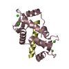
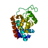
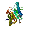
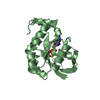
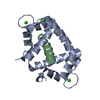
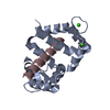
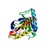
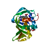
- PDB-1vrk: THE 1.9 ANGSTROM STRUCTURE OF E84K-CALMODULIN RS20 PEPTIDE COMPLEX -
+ Open data

- Basic information
| Entry | Database: PDB / ID: 1vrk | ||||||
|---|---|---|---|---|---|---|---|
| Title | THE 1.9 ANGSTROM STRUCTURE OF E84K-CALMODULIN RS20 PEPTIDE COMPLEX | ||||||
 Components Components |
| ||||||
 Keywords Keywords | COMPLEX(CALCIUM-BINDING PROTEIN/PEPTIDE) / CALMODULIN / CALCIUM BINDING / HELIX-LOOP-HELIX / SIGNALLING / COMPLEX(CALCIUM-BINDING PROTEIN-PEPTIDE) / COMPLEX(CALCIUM-BINDING PROTEIN-PEPTIDE) complex | ||||||
| Function / homology |  Function and homology information Function and homology informationtonic smooth muscle contraction / myosin-light-chain kinase / myosin light chain kinase activity / positive regulation of sarcomere organization / positive regulation of striated muscle contraction / cardiac muscle cell development / cleavage furrow / heart morphogenesis / stress fiber / lamellipodium ...tonic smooth muscle contraction / myosin-light-chain kinase / myosin light chain kinase activity / positive regulation of sarcomere organization / positive regulation of striated muscle contraction / cardiac muscle cell development / cleavage furrow / heart morphogenesis / stress fiber / lamellipodium / calmodulin binding / ATP binding / metal ion binding / cytosol / cytoplasm Similarity search - Function | ||||||
| Biological species | synthetic construct (others) | ||||||
| Method |  X-RAY DIFFRACTION / MOLECULAR REPLACEMENT / Resolution: 1.9 Å X-RAY DIFFRACTION / MOLECULAR REPLACEMENT / Resolution: 1.9 Å | ||||||
 Authors Authors | Weigand, S. / Anderson, W.F. | ||||||
 Citation Citation | Journal: Biochemistry / Year: 1999 Title: Analysis of the functional coupling between calmodulin's calcium binding and peptide recognition properties. Authors: Mirzoeva, S. / Weigand, S. / Lukas, T.J. / Shuvalova, L. / Anderson, W.F. / Watterson, D.M. #1: Journal: Science / Year: 1992Title: Target Enzyme Recognition by Calmodulin: 2.4 A Structure of a Calmodulin-Peptide Complex Authors: Meador, W.E. / Means, A.R. / Quiocho, F.A. | ||||||
| History |
| ||||||
| Remark 650 | HELIX THE STRUCTURAL CONSEQUENCE OF THE E84K MUTATION RELATIVE TO THE WILD TYPE STRUCTURE ...HELIX THE STRUCTURAL CONSEQUENCE OF THE E84K MUTATION RELATIVE TO THE WILD TYPE STRUCTURE (REFERENCE 1 ABOVE) IS AN ALTERATION OF THE RESIDUE 84 CONFORMATION AND A FIVE DEGREE MOVEMENT OF HELIX E (HELIX 5) AWAY FROM THE PEPTIDE |
- Structure visualization
| Structure viewer | Molecule: MolmilJmol/JSmol |
|---|
- Downloads & links
-Download
| PDBx/mmCIF format | 1vrk.cif.gz | 53.1 KB | Display | PDBx/mmCIF format |
|---|---|---|---|---|
| PDB format | pdb1vrk.ent.gz | 36.9 KB | Display | PDB format |
| PDBx/mmJSON format | 1vrk.json.gz | Tree view | PDBx/mmJSON format | |
| Others |  Other downloads Other downloads |
-Validation report
| Arichive directory | https://data.pdbj.org/pub/pdb/validation_reports/vr/1vrkftp://data.pdbj.org/pub/pdb/validation_reports/vr/1vrk | HTTPS FTP |
|---|
-Related structure data
| Related structure data | 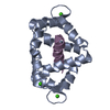 1cdlS S: Starting model for refinement |
|---|---|
| Similar structure data |


-Links
 PDBj
PDBj


- Assembly
| Deposited unit | 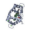
| ||||||||
|---|---|---|---|---|---|---|---|---|---|
| 1 |
| ||||||||
| Unit cell |
|
-Components
| #1: Protein | Mass: 16642.338 Da / Num. of mol.: 1 / Mutation: E84K Source method: isolated from a genetically manipulated source Source: (gene. exp.) synthetic construct (others) / Description: CONSENSUS SEQUENCE FROM A NUMBER OF SOURCES; / Plasmid: PVUCH-1 / Production host:  | ||||||||
|---|---|---|---|---|---|---|---|---|---|
| #2: Protein/peptide | Mass: 2327.696 Da / Num. of mol.: 1 Source method: isolated from a genetically manipulated source Details: TRP 4 IS NE-FORMYLATED / References: UniProt: P11799*PLUS, EC: 2.7.1.117 | ||||||||
| #3: Chemical | ChemComp-CA /   Mass: 40.078 Da / Num. of mol.: 4 / Source method: obtained synthetically / Formula: Ca Mass: 40.078 Da / Num. of mol.: 4 / Source method: obtained synthetically / Formula: Ca#4: Chemical | ChemComp-ACT / |   Mass: 59.044 Da / Num. of mol.: 1 / Source method: obtained synthetically / Formula: C2H3O2 Mass: 59.044 Da / Num. of mol.: 1 / Source method: obtained synthetically / Formula: C2H3O2#5: Water | ChemComp-HOH / |  Mass: 18.015 Da / Num. of mol.: 141 / Source method: isolated from a natural source / Formula: H2O Mass: 18.015 Da / Num. of mol.: 141 / Source method: isolated from a natural source / Formula: H2OCompound details | THE ENHANCEMENT OF CALMODULIN'S CALCIUM BINDING BY SMOOTH MUSCLE/NONMUSCLE MYOSIN LIGHT CHAIN ...THE ENHANCEMEN | Has protein modification | Y | |
-Experimental details
-Experiment
| Experiment | Method: X-RAY DIFFRACTION / Number of used crystals: 2 |
|---|
- Sample preparation
| Crystal | Density Matthews: 2.1 Å3/Da / Density % sol: 43 % | ||||||||||||||||||||||||||||||||||||||||||||||||||||||||||||||||||||||||
|---|---|---|---|---|---|---|---|---|---|---|---|---|---|---|---|---|---|---|---|---|---|---|---|---|---|---|---|---|---|---|---|---|---|---|---|---|---|---|---|---|---|---|---|---|---|---|---|---|---|---|---|---|---|---|---|---|---|---|---|---|---|---|---|---|---|---|---|---|---|---|---|---|---|
| Crystal grow | pH: 4.6 / Details: pH 4.6 | ||||||||||||||||||||||||||||||||||||||||||||||||||||||||||||||||||||||||
| Crystal | *PLUS | ||||||||||||||||||||||||||||||||||||||||||||||||||||||||||||||||||||||||
| Crystal grow | *PLUS Temperature: 4 ℃ / pH: 7.5 / Method: vapor diffusion, hanging drop | ||||||||||||||||||||||||||||||||||||||||||||||||||||||||||||||||||||||||
| Components of the solutions | *PLUS
|
-Data collection
| Diffraction | Mean temperature: 293 K |
|---|---|
| Diffraction source | Source: ROTATING ANODE / Type: RIGAKU RUH2R / Wavelength: 1.5418 |
| Detector | Type: XUONG-HAMLIN MULTIWIRE / Detector: AREA DETECTOR / Date: Apr 1, 1993 / Details: COLLIMATOR |
| Radiation | Monochromator: GRAPHITE(002) / Monochromatic (M) / Laue (L): M / Scattering type: x-ray |
| Radiation wavelength | Wavelength: 1.5418 Å / Relative weight: 1 |
| Reflection | Resolution: 1.9→18.2 Å / Num. obs: 11190 / % possible obs: 88 % / Observed criterion σ(I): 0 / Redundancy: 3.2 % / Biso Wilson estimate: 7.6 Å2 / Rmerge(I) obs: 0.0649 / Net I/σ(I): 15 |
| Reflection shell | Resolution: 1.9→2.05 Å / Redundancy: 1.9 % / Rmerge(I) obs: 0.237 / Mean I/σ(I) obs: 2 / % possible all: 65 |
- Processing
| Software |
| ||||||||||||||||||||||||||||||||||||||||||||||||||||||||||||||||||||||||||||||||
|---|---|---|---|---|---|---|---|---|---|---|---|---|---|---|---|---|---|---|---|---|---|---|---|---|---|---|---|---|---|---|---|---|---|---|---|---|---|---|---|---|---|---|---|---|---|---|---|---|---|---|---|---|---|---|---|---|---|---|---|---|---|---|---|---|---|---|---|---|---|---|---|---|---|---|---|---|---|---|---|---|---|
| Refinement | Method to determine structure: MOLECULAR REPLACEMENT Starting model: PDB ENTRY 1CDL: SECOND COMPLEX (SEGID B AND F) OF THE ASYMMETRIC UNIT Resolution: 1.9→30 Å / Rfactor Rfree error: 0.008 / Data cutoff high absF: 10000000 / Data cutoff low absF: 0.001 / Isotropic thermal model: RESTRAINED / Cross valid method: THROUGHOUT / σ(F): 2 / Details: BULK SOLVENT MODEL USED
| ||||||||||||||||||||||||||||||||||||||||||||||||||||||||||||||||||||||||||||||||
| Displacement parameters | Biso mean: 30.7 Å2 | ||||||||||||||||||||||||||||||||||||||||||||||||||||||||||||||||||||||||||||||||
| Refine analyze |
| ||||||||||||||||||||||||||||||||||||||||||||||||||||||||||||||||||||||||||||||||
| Refinement step | Cycle: LAST / Resolution: 1.9→30 Å
| ||||||||||||||||||||||||||||||||||||||||||||||||||||||||||||||||||||||||||||||||
| Refine LS restraints |
| ||||||||||||||||||||||||||||||||||||||||||||||||||||||||||||||||||||||||||||||||
| LS refinement shell | Resolution: 1.9→2.02 Å / Rfactor Rfree error: 0.035 / Total num. of bins used: 6
| ||||||||||||||||||||||||||||||||||||||||||||||||||||||||||||||||||||||||||||||||
| Xplor file |
| ||||||||||||||||||||||||||||||||||||||||||||||||||||||||||||||||||||||||||||||||
| Software | *PLUS Name: X-PLOR / Version: 3.851 / Classification: refinement | ||||||||||||||||||||||||||||||||||||||||||||||||||||||||||||||||||||||||||||||||
| Refine LS restraints | *PLUS
|
