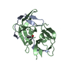
endochitinase activity / chitinase / chitin catabolic process / chitin binding / polysaccharide catabolic process / defense response to fungus / cell wall macromolecule catabolic process / killing of cells of another organism / extracellular region Similarity search - Function
Chitinases family 19 signature 1. / Endochitinase; domain 2 / Endochitinase, domain 2 / Chitinases family 19 signature 2. / Glycoside hydrolase, family 19 / Glycoside hydrolase, family 19, catalytic / Chitinase class I / Chitin-binding, type 1, conserved site / Chitin recognition or binding domain signature. / Chitin-binding type-1 domain profile. ...Chitinases family 19 signature 1. / Endochitinase; domain 2 / Endochitinase, domain 2 / Chitinases family 19 signature 2. / Glycoside hydrolase, family 19 / Glycoside hydrolase, family 19, catalytic / Chitinase class I / Chitin-binding, type 1, conserved site / Chitin recognition or binding domain signature. / Chitin-binding type-1 domain profile. / Chitin binding domain / Chitin-binding, type 1 / Endochitinase-like superfamily / Lysozyme - #10 / Lysozyme / Lysozyme-like domain superfamily / 2-Layer Sandwich / Orthogonal Bundle / Mainly Alpha / Alpha Beta Similarity search - Domain/homology
 Movie
Movie Controller

 Yorodumi
 Open data
 Basic information
 Components
 Keywords
 Function and homology information
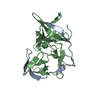
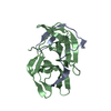
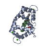
 Cryptomeria japonica (Japanese cedar)
 X-RAY DIFFRACTION /
  Authors
 Citation
 Structure visualization
 Downloads & links
 Other downloads


 PDBj
PDBj Assembly
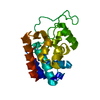

 Mass: 18.015 Da / Num. of mol.: 183 / Source method: isolated from a natural source / Formula: H2O
 Sample preparation
 / Beamline: BL-17A / Wavelength: 0.98 Å
 Processing