 Movie
Movie Controller

[English] 日本語
 Yorodumi
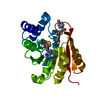
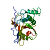
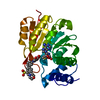
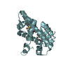
- PDB-4dr9: Crystal structure of a peptide deformylase from synechococcus elo... -
+ Open data

- Basic information
| Entry | Database: PDB / ID: 4dr9 | ||||||
|---|---|---|---|---|---|---|---|
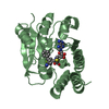
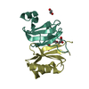
| Title | Crystal structure of a peptide deformylase from synechococcus elongatus in complex with actinonin | ||||||
 Components Components | Peptide deformylase | ||||||
 Keywords Keywords | HYDROLASE/HYDROLASE INHIBITOR / HYDROLASE-HYDROLASE INHIBITOR complex | ||||||
| Function / homology |  Function and homology information Function and homology informationpeptide deformylase / peptide deformylase activity / translation / metal ion binding Similarity search - Function | ||||||
| Biological species |  Synechococcus elongatus (bacteria) Synechococcus elongatus (bacteria) | ||||||
| Method |  X-RAY DIFFRACTION / SYNCHROTRON / MOLECULAR REPLACEMENT / Resolution: 1.9 Å X-RAY DIFFRACTION / SYNCHROTRON / MOLECULAR REPLACEMENT / Resolution: 1.9 Å | ||||||
 Authors Authors | Lorimer, D. / Abendroth, J. / Craig, T. / Burgin, A. / Segall, A. / Rohwler, F. | ||||||
 Citation Citation | Journal: ISME J / Year: 2013 Title: Structure and function of a cyanophage-encoded peptide deformylase. Authors: Frank, J.A. / Lorimer, D. / Youle, M. / Witte, P. / Craig, T. / Abendroth, J. / Rohwer, F. / Edwards, R.A. / Segall, A.M. / Burgin, A.B. | ||||||
| History |
|
- Structure visualization
| Structure viewer | Molecule: MolmilJmol/JSmol |
|---|
- Downloads & links
-Download
| PDBx/mmCIF format | 4dr9.cif.gz | 302.5 KB | Display | PDBx/mmCIF format |
|---|---|---|---|---|
| PDB format | pdb4dr9.ent.gz | 247.3 KB | Display | PDB format |
| PDBx/mmJSON format | 4dr9.json.gz | Tree view | PDBx/mmJSON format | |
| Others |  Other downloads Other downloads |
-Validation report
| Arichive directory | https://data.pdbj.org/pub/pdb/validation_reports/dr/4dr9ftp://data.pdbj.org/pub/pdb/validation_reports/dr/4dr9 | HTTPS FTP |
|---|
-Related structure data
| Related structure data |  3uwaC  3uwbC  4dr8C 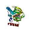 1lryS C: citing same article ( S: Starting model for refinement |
|---|---|
| Similar structure data |


-Links
 PDBj
PDBj

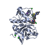
- Assembly
| Deposited unit | 
| ||||||||
|---|---|---|---|---|---|---|---|---|---|
| 1 | 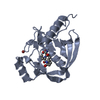
| ||||||||
| 2 | 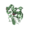
| ||||||||
| 3 | 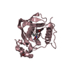
| ||||||||
| 4 | 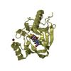
| ||||||||
| Unit cell |
|
-Components
| #1: Protein | Mass: 21333.562 Da / Num. of mol.: 4 Source method: isolated from a genetically manipulated source Source: (gene. exp.) Synechococcus elongatus (bacteria) / Strain: PCC6301 / Gene: def, syc0213_d, YP_170923 / Plasmid: VCID 6622 / Production host: References: UniProt: Q5N5L5, UniProt: A0A0H3JZJ4*PLUS, peptide deformylase #2: Chemical | ChemComp-ZN /   Mass: 65.409 Da / Num. of mol.: 4 / Source method: obtained synthetically / Formula: Zn Mass: 65.409 Da / Num. of mol.: 4 / Source method: obtained synthetically / Formula: Zn#3: Chemical | ChemComp-BR /   Mass: 79.904 Da / Num. of mol.: 5 / Source method: obtained synthetically / Formula: Br Mass: 79.904 Da / Num. of mol.: 5 / Source method: obtained synthetically / Formula: Br#4: Chemical | ChemComp-BB2 /   Mass: 385.498 Da / Num. of mol.: 4 / Source method: obtained synthetically / Formula: C19H35N3O5 / Comment: antibiotic*YM Mass: 385.498 Da / Num. of mol.: 4 / Source method: obtained synthetically / Formula: C19H35N3O5 / Comment: antibiotic*YM#5: Water | ChemComp-HOH / |  Mass: 18.015 Da / Num. of mol.: 564 / Source method: isolated from a natural source / Formula: H2O Mass: 18.015 Da / Num. of mol.: 564 / Source method: isolated from a natural source / Formula: H2O |
|---|
-Experimental details
-Experiment
| Experiment | Method: X-RAY DIFFRACTION / Number of used crystals: 1 |
|---|
- Sample preparation
| Crystal | Density Matthews: 2.19 Å3/Da / Density % sol: 43.9 % |
|---|---|
| Crystal grow | Temperature: 290 K / Method: vapor diffusion, sitting drop / pH: 7.5 Details: HAMPTON RESEARCH INDEX H12: 30% PEG 2000 MME, 150MM KBR, PH 7.5, VAPOR DIFFUSION, SITTING DROP, TEMPERATURE 290K; 18 hour soak in reservoir solution containing 1mM actinonin |
-Data collection
| Diffraction |
| ||||||||||||||||||
|---|---|---|---|---|---|---|---|---|---|---|---|---|---|---|---|---|---|---|---|
| Diffraction source |
| ||||||||||||||||||
| Detector | Type: RIGAKU SATURN 944+ / Detector: CCD / Date: Feb 7, 2012 / Details: RIGAKU VARIMAX | ||||||||||||||||||
| Radiation | Monochromator: RIGAKU VARIMAX / Protocol: SINGLE WAVELENGTH / Monochromatic (M) / Laue (L): M / Scattering type: x-ray | ||||||||||||||||||
| Radiation wavelength |
| ||||||||||||||||||
| Reflection | Resolution: 1.9→50 Å / Num. all: 56808 / Num. obs: 56579 / % possible obs: 99.6 % / Observed criterion σ(F): 0 / Observed criterion σ(I): -3 / Redundancy: 5.8 % / Biso Wilson estimate: 22.89 Å2 / Rmerge(I) obs: 0.077 / Rsym value: 0.077 / Net I/σ(I): 17 | ||||||||||||||||||
| Reflection shell | Resolution: 1.9→1.95 Å / Redundancy: 2.5 % / Rmerge(I) obs: 0.49 / Mean I/σ(I) obs: 2.02 / Num. unique all: 4200 / Rsym value: 0.49 / % possible all: 98 |

- Processing
| Software |
| ||||||||||||||||||||||||||||||||||||||||||||||||||||||||||||||||||||||||||||||||||||||||||||||||||||||||||||||||||||||||||||||||||||||||||||||||||||||||||||||||||||||||||
|---|---|---|---|---|---|---|---|---|---|---|---|---|---|---|---|---|---|---|---|---|---|---|---|---|---|---|---|---|---|---|---|---|---|---|---|---|---|---|---|---|---|---|---|---|---|---|---|---|---|---|---|---|---|---|---|---|---|---|---|---|---|---|---|---|---|---|---|---|---|---|---|---|---|---|---|---|---|---|---|---|---|---|---|---|---|---|---|---|---|---|---|---|---|---|---|---|---|---|---|---|---|---|---|---|---|---|---|---|---|---|---|---|---|---|---|---|---|---|---|---|---|---|---|---|---|---|---|---|---|---|---|---|---|---|---|---|---|---|---|---|---|---|---|---|---|---|---|---|---|---|---|---|---|---|---|---|---|---|---|---|---|---|---|---|---|---|---|---|---|---|---|
| Refinement | Method to determine structure: MOLECULAR REPLACEMENT Starting model: pdb deposition 1lry, modified with CCP4 program CHAINSAW Resolution: 1.9→50 Å / Cor.coef. Fo:Fc: 0.949 / Cor.coef. Fo:Fc free: 0.913 / SU B: 6.776 / SU ML: 0.104 / Isotropic thermal model: isotropic, TLS / Cross valid method: THROUGHOUT / σ(F): 0 / ESU R: 0.166 / ESU R Free: 0.156 / Stereochemistry target values: MAXIMUM LIKELIHOOD / Details: HYDROGENS HAVE BEEN ADDED IN THE
| ||||||||||||||||||||||||||||||||||||||||||||||||||||||||||||||||||||||||||||||||||||||||||||||||||||||||||||||||||||||||||||||||||||||||||||||||||||||||||||||||||||||||||
| Solvent computation | Ion probe radii: 0.8 Å / Shrinkage radii: 0.8 Å / VDW probe radii: 1.2 Å / Solvent model: MASK | ||||||||||||||||||||||||||||||||||||||||||||||||||||||||||||||||||||||||||||||||||||||||||||||||||||||||||||||||||||||||||||||||||||||||||||||||||||||||||||||||||||||||||
| Displacement parameters | Biso mean: 18.66 Å2
| ||||||||||||||||||||||||||||||||||||||||||||||||||||||||||||||||||||||||||||||||||||||||||||||||||||||||||||||||||||||||||||||||||||||||||||||||||||||||||||||||||||||||||
| Refinement step | Cycle: LAST / Resolution: 1.9→50 Å
| ||||||||||||||||||||||||||||||||||||||||||||||||||||||||||||||||||||||||||||||||||||||||||||||||||||||||||||||||||||||||||||||||||||||||||||||||||||||||||||||||||||||||||
| Refine LS restraints |
| ||||||||||||||||||||||||||||||||||||||||||||||||||||||||||||||||||||||||||||||||||||||||||||||||||||||||||||||||||||||||||||||||||||||||||||||||||||||||||||||||||||||||||
| LS refinement shell | Resolution: 1.9→1.95 Å / Total num. of bins used: 20
| ||||||||||||||||||||||||||||||||||||||||||||||||||||||||||||||||||||||||||||||||||||||||||||||||||||||||||||||||||||||||||||||||||||||||||||||||||||||||||||||||||||||||||
| Refinement TLS params. | Method: refined / Refine-ID: X-RAY DIFFRACTION
| ||||||||||||||||||||||||||||||||||||||||||||||||||||||||||||||||||||||||||||||||||||||||||||||||||||||||||||||||||||||||||||||||||||||||||||||||||||||||||||||||||||||||||
| Refinement TLS group |
|
