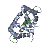
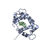
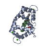
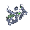
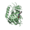
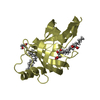
[English] 日本語
Yorodumi
- PDB-4q5u: Structure of calmodulin bound to its recognition site from calcineurin -

+
Open data


ID or keywords:

Loading...

-
Basic information

Entry
Database: PDB / ID: 4q5u
TitleStructure of calmodulin bound to its recognition site from calcineurin
Components
  • Calmodulin
  • Serine/threonine-protein phosphatase 2B catalytic subunit alpha isoform
KeywordsCALCIUM BINDING PROTEIN/PROTEIN BINDING / EF hand / CALCIUM BINDING PROTEIN-PROTEIN BINDING complex
Function / homology
Function and homology information


negative regulation of angiotensin-activated signaling pathway / regulation of cell proliferation involved in kidney morphogenesis / positive regulation of glomerulus development / negative regulation of calcium ion import across plasma membrane / protein serine/threonine phosphatase complex / negative regulation of signaling / positive regulation of saliva secretion / positive regulation of cardiac muscle hypertrophy in response to stress / positive regulation of calcium ion import across plasma membrane / calmodulin-dependent protein phosphatase activity ...negative regulation of angiotensin-activated signaling pathway / regulation of cell proliferation involved in kidney morphogenesis / positive regulation of glomerulus development / negative regulation of calcium ion import across plasma membrane / protein serine/threonine phosphatase complex / negative regulation of signaling / positive regulation of saliva secretion / positive regulation of cardiac muscle hypertrophy in response to stress / positive regulation of calcium ion import across plasma membrane / calmodulin-dependent protein phosphatase activity / calcineurin complex / positive regulation of connective tissue replacement / negative regulation of dendrite morphogenesis / calcineurin-mediated signaling / slit diaphragm / peptidyl-serine dephosphorylation / calcineurin-NFAT signaling cascade / renal filtration / skeletal muscle tissue regeneration / : / transition between fast and slow fiber / positive regulation of calcineurin-NFAT signaling cascade / establishment of protein localization to mitochondrial membrane / type 3 metabotropic glutamate receptor binding / positive regulation of osteoclast differentiation / cardiac muscle hypertrophy in response to stress / CaM pathway / Cam-PDE 1 activation / dendrite morphogenesis / Sodium/Calcium exchangers / regulation of synaptic vesicle endocytosis / Calmodulin induced events / Reduction of cytosolic Ca++ levels / CREB1 phosphorylation through the activation of CaMKII/CaMKK/CaMKIV cascasde / Activation of Ca-permeable Kainate Receptor / Loss of phosphorylation of MECP2 at T308 / CREB1 phosphorylation through the activation of Adenylate Cyclase / PKA activation / negative regulation of high voltage-gated calcium channel activity / CaMK IV-mediated phosphorylation of CREB / Glycogen breakdown (glycogenolysis) / cyclosporin A binding / positive regulation of cyclic-nucleotide phosphodiesterase activity / organelle localization by membrane tethering / negative regulation of calcium ion export across plasma membrane / regulation of synaptic vesicle exocytosis / myosin phosphatase activity / CLEC7A (Dectin-1) induces NFAT activation / regulation of cardiac muscle cell action potential / mitochondrion-endoplasmic reticulum membrane tethering / autophagosome membrane docking / Activation of RAC1 downstream of NMDARs / postsynaptic modulation of chemical synaptic transmission / extrinsic component of plasma membrane / response to corticosterone / protein serine/threonine phosphatase activity / protein-serine/threonine phosphatase / nitric-oxide synthase binding / positive regulation of ryanodine-sensitive calcium-release channel activity / positive regulation of activated T cell proliferation / regulation of cell communication by electrical coupling involved in cardiac conduction / Negative regulation of NMDA receptor-mediated neuronal transmission / negative regulation of peptidyl-threonine phosphorylation / Synthesis of IP3 and IP4 in the cytosol / Unblocking of NMDA receptors, glutamate binding and activation / Phase 0 - rapid depolarisation / protein phosphatase activator activity / RHO GTPases activate PAKs / positive regulation of endocytosis / positive regulation of phosphoprotein phosphatase activity / Ion transport by P-type ATPases / Long-term potentiation / Uptake and function of anthrax toxins / Calcineurin activates NFAT / Regulation of MECP2 expression and activity / adenylate cyclase binding / catalytic complex / positive regulation of cell adhesion / DARPP-32 events / detection of calcium ion / regulation of cardiac muscle contraction / negative regulation of ryanodine-sensitive calcium-release channel activity / Smooth Muscle Contraction / epidermis development / RHO GTPases activate IQGAPs / calcium channel inhibitor activity / cellular response to interferon-beta / positive regulation of DNA binding / regulation of cardiac muscle contraction by regulation of the release of sequestered calcium ion / negative regulation of insulin secretion / Protein methylation / multicellular organismal response to stress / positive regulation of osteoblast differentiation / phosphatidylinositol 3-kinase binding / eNOS activation / skeletal muscle fiber development / Activation of AMPK downstream of NMDARs / regulation of release of sequestered calcium ion into cytosol by sarcoplasmic reticulum / enzyme regulator activity / dephosphorylation
Similarity search - Function
PP2B, metallophosphatase domain / PP2B / Serine/threonine specific protein phosphatases signature. / Protein phosphatase 2A homologues, catalytic domain. / Serine/threonine-specific protein phosphatase/bis(5-nucleosyl)-tetraphosphatase / Calcineurin-like phosphoesterase domain, ApaH type / Calcineurin-like phosphoesterase / Metallo-dependent phosphatase-like / EF-hand domain pair / EF-hand, calcium binding motif ...PP2B, metallophosphatase domain / PP2B / Serine/threonine specific protein phosphatases signature. / Protein phosphatase 2A homologues, catalytic domain. / Serine/threonine-specific protein phosphatase/bis(5-nucleosyl)-tetraphosphatase / Calcineurin-like phosphoesterase domain, ApaH type / Calcineurin-like phosphoesterase / Metallo-dependent phosphatase-like / EF-hand domain pair / EF-hand, calcium binding motif / EF-Hand 1, calcium-binding site / EF-hand calcium-binding domain. / EF-hand calcium-binding domain profile. / EF-hand domain / EF-hand domain pair
Similarity search - Domain/homology
Calmodulin-1 / Calmodulin-3 / Protein phosphatase 3 catalytic subunit alpha
Similarity search - Component
Biological speciesHomo sapiens (human)
MethodX-RAY DIFFRACTION / SYNCHROTRON / MOLECULAR REPLACEMENT / Resolution: 1.95 Å
AuthorsGuo, H. / Dunlap, T.B. / Creamer, T.P. / Vander Kooi, C.W.
CitationJournal: Biochemistry / Year: 2014
Title: Stoichiometry of the calcineurin regulatory domain-calmodulin complex.
Authors: Dunlap, T.B. / Guo, H.F. / Cook, E.C. / Holbrook, E. / Rumi-Masante, J. / Lester, T.E. / Colbert, C.L. / Vander Kooi, C.W. / Creamer, T.P.
History
DepositionApr 17, 2014Deposition site: RCSB / Processing site: RCSB
Revision 1.0Sep 3, 2014Provider: repository / Type: Initial release
Revision 1.1Oct 1, 2014Group: Database references
Revision 1.2Sep 20, 2023Group: Data collection / Database references ...Data collection / Database references / Derived calculations / Refinement description
Category: chem_comp_atom / chem_comp_bond ...chem_comp_atom / chem_comp_bond / database_2 / pdbx_initial_refinement_model / pdbx_struct_conn_angle / struct_conn / struct_site
Item: _database_2.pdbx_DOI / _database_2.pdbx_database_accession ..._database_2.pdbx_DOI / _database_2.pdbx_database_accession / _pdbx_struct_conn_angle.ptnr1_auth_comp_id / _pdbx_struct_conn_angle.ptnr1_auth_seq_id / _pdbx_struct_conn_angle.ptnr1_label_asym_id / _pdbx_struct_conn_angle.ptnr1_label_atom_id / _pdbx_struct_conn_angle.ptnr1_label_comp_id / _pdbx_struct_conn_angle.ptnr1_label_seq_id / _pdbx_struct_conn_angle.ptnr2_auth_seq_id / _pdbx_struct_conn_angle.ptnr2_label_asym_id / _pdbx_struct_conn_angle.ptnr3_auth_comp_id / _pdbx_struct_conn_angle.ptnr3_auth_seq_id / _pdbx_struct_conn_angle.ptnr3_label_asym_id / _pdbx_struct_conn_angle.ptnr3_label_atom_id / _pdbx_struct_conn_angle.ptnr3_label_comp_id / _pdbx_struct_conn_angle.ptnr3_label_seq_id / _pdbx_struct_conn_angle.value / _struct_conn.pdbx_dist_value / _struct_conn.ptnr1_auth_comp_id / _struct_conn.ptnr1_auth_seq_id / _struct_conn.ptnr1_label_asym_id / _struct_conn.ptnr1_label_atom_id / _struct_conn.ptnr1_label_comp_id / _struct_conn.ptnr1_label_seq_id / _struct_conn.ptnr2_auth_seq_id / _struct_conn.ptnr2_label_asym_id / _struct_site.pdbx_auth_asym_id / _struct_site.pdbx_auth_comp_id / _struct_site.pdbx_auth_seq_id

-
Structure visualization

Structure viewerMolecule:
MolmilJmol/JSmol

Downloads & links

-
Assembly

Deposited unit
A: Calmodulin
C: Serine/threonine-protein phosphatase 2B catalytic subunit alpha isoform
hetero molecules


Theoretical massNumber of molelcules
Total (without water)19,8336
Polymers19,6732
Non-polymers1604
Water1,802100
1


  • Idetical with deposited unit
  • defined by author&software
TypeNameSymmetry operationNumber
identity operation1_555x,y,z1
Buried area3280 Å2
ΔGint-74 kcal/mol
Surface area9050 Å2
MethodPISA
Unit cell
Length a, b, c (Å)105.499, 111.033, 39.448
Angle α, β, γ (deg.)90.00, 90.00, 90.00
Int Tables number20
Space group name H-MC2221
Components on special symmetry positions
IDModelComponents
11A-340-

HOH

-
Components

#1: Protein Calmodulin / CaM


Mass: 16852.545 Da / Num. of mol.: 1
Source method: isolated from a genetically manipulated source
Source: (gene. exp.) Homo sapiens (human)
Gene: CALM1, CALM, CAM, CAM1, CALM2, CAM2, CAMB, CALM3, CALML2, CAM3, CAMC, CAMIII
Plasmid: pETCaMI / Production host: Escherichia coli (E. coli) / Strain (production host): BL21(DE3) / References: UniProt: P62158, UniProt: P0DP23*PLUS
#2: Protein/peptide Serine/threonine-protein phosphatase 2B catalytic subunit alpha isoform / CAM-PRP catalytic subunit / Calmodulin-dependent calcineurin A subunit alpha isoform


Mass: 2820.496 Da / Num. of mol.: 1 / Fragment: calmodulin-binding domain (UNP residues 391-414) / Source method: obtained synthetically / Source: (synth.) Homo sapiens (human) / References: UniProt: Q08209
#3: Chemical
ChemComp-CA / CALCIUM ION


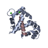
Mass: 40.078 Da / Num. of mol.: 4 / Source method: obtained synthetically / Formula: Ca
#4: Water ChemComp-HOH / water


Mass: 18.015 Da / Num. of mol.: 100 / Source method: isolated from a natural source / Formula: H2O

-
Experimental details

-
Experiment

ExperimentMethod: X-RAY DIFFRACTION / Number of used crystals: 1

-
Sample preparation

CrystalDensity Matthews: 2.94 Å3/Da / Density % sol: 58.11 %
Crystal growTemperature: 298 K / Method: vapor diffusion, hanging drop / pH: 7.5
Details: 3:1 10 mg/ml protein to mother liquor (24% PEG1000, 20% glycerol), final volume 200 nL, pH 7.5, VAPOR DIFFUSION, HANGING DROP, temperature 298.0K

-
Data collection

DiffractionMean temperature: 100 K
Diffraction sourceSource: SYNCHROTRON / Site: APS / Beamline: 22-ID / Wavelength: 1 Å
DetectorType: MARMOSAIC 300 mm CCD / Detector: CCD / Date: Aug 17, 2011
RadiationMonochromator: Rosenbaum-Rock double-crystal Si(111) / Protocol: SINGLE WAVELENGTH / Monochromatic (M) / Laue (L): M / Scattering type: x-ray
Radiation wavelengthWavelength: 1 Å / Relative weight: 1
ReflectionResolution: 1.95→19.12 Å / Num. obs: 15779 / % possible obs: 91.1 % / Observed criterion σ(I): -3 / Redundancy: 5.5 % / Rmerge(I) obs: 0.098 / Net I/σ(I): 14.4
Reflection shellResolution: 1.95→2.02 Å / Redundancy: 3.2 % / Rmerge(I) obs: 0.375 / Mean I/σ(I) obs: 3.4 / % possible all: 69.6

-
Processing

Software
NameVersionClassification
HKL-2000data collection
PHASERphasing
PHENIX(phenix.refine: 1.8_1069)refinement
HKL-2000data reduction
HKL-2000data scaling
RefinementMethod to determine structure: MOLECULAR REPLACEMENT
Starting model: PDB ENTRY 2W73
Resolution: 1.95→19.12 Å / SU ML: 0.17 / σ(F): 1.38 / Phase error: 30.04 / Stereochemistry target values: ML
RfactorNum. reflection% reflectionSelection details
Rfree0.2484 801 5.08 %RANDOM
Rwork0.2147 ---
obs0.2165 15779 90.91 %-
Solvent computationShrinkage radii: 0.9 Å / VDW probe radii: 1.11 Å / Solvent model: FLAT BULK SOLVENT MODEL
Refinement stepCycle: LAST / Resolution: 1.95→19.12 Å
ProteinNucleic acidLigandSolventTotal
Num. atoms1340 0 4 100 1444
Refine LS restraints
Refine-IDTypeDev idealNumber
X-RAY DIFFRACTIONf_bond_d0.0061352
X-RAY DIFFRACTIONf_angle_d0.9721810
X-RAY DIFFRACTIONf_dihedral_angle_d14.43524
X-RAY DIFFRACTIONf_chiral_restr0.07200
X-RAY DIFFRACTIONf_plane_restr0.004244
LS refinement shell
Resolution (Å)Rfactor RfreeNum. reflection RfreeRfactor RworkNum. reflection RworkRefine-ID% reflection obs (%)
1.95-2.07160.3421000.27111902X-RAY DIFFRACTION70
2.0716-2.23130.29661110.24722211X-RAY DIFFRACTION82
2.2313-2.45550.26081450.23642531X-RAY DIFFRACTION93
2.4555-2.80980.27241420.23142714X-RAY DIFFRACTION100
2.8098-3.53640.22951510.22812749X-RAY DIFFRACTION100
3.5364-19.12110.23541520.19142871X-RAY DIFFRACTION100
Refinement TLS params.

Method: refined / Refine-ID: X-RAY DIFFRACTION

IDL112)L122)L132)L222)L232)L332)S11 (Å °)S12 (Å °)S13 (Å °)S21 (Å °)S22 (Å °)S23 (Å °)S31 (Å °)S32 (Å °)S33 (Å °)T112)T122)T132)T222)T232)T332)Origin x (Å)Origin y (Å)Origin z (Å)
13.8011-0.6887-2.58268.36052.72684.4845-0.1732-0.0153-0.2597-0.1124-0.05150.0840.40120.05830.31390.45810.0503-0.05650.4150.02590.440732.773610.21436.8774
25.6530.0406-0.28182.76-0.31999.32380.2090.2182-0.20680.0644-0.36180.10890.9835-0.3170.19750.50420.0595-0.11190.4474-0.01140.506931.215322.8885-4.3999
34.47940.1059-0.35384.9089-3.92523.343-0.0861-0.14670.59880.2157-0.8049-1.376-0.40581.7490.73220.3862-0.0185-0.09140.77420.10010.920645.258228.1702-1.9114
49.989-2.274-1.8637.8131.08187.19950.0810.4257-0.0722-0.3109-0.2869-0.13530.28940.00880.07760.46680.016-0.10250.42210.02150.426934.831917.7501-0.2849
Refinement TLS group
IDRefine-IDRefine TLS-IDSelection details
1X-RAY DIFFRACTION1(chain A and resid 3:73)
2X-RAY DIFFRACTION2(chain A and resid 74:97)
3X-RAY DIFFRACTION3(chain A and resid 98:147)
4X-RAY DIFFRACTION4(chain C and resid 391:414)

+
About Yorodumi

-
News

-
Feb 9, 2022. New format data for meta-information of EMDB entries

New format data for meta-information of EMDB entries

  • Version 3 of the EMDB header file is now the official format.
  • The previous official version 1.9 will be removed from the archive.

Related info.:EMDB header

External links:wwPDB to switch to version 3 of the EMDB data model

-
Aug 12, 2020. Covid-19 info

Covid-19 info

URL: https://pdbjlvh1.pdbj.org/emnavi/covid19.php

New page: Covid-19 featured information page in EM Navigator.

Related info.:Covid-19 info / Mar 5, 2020. Novel coronavirus structure data

+
Mar 5, 2020. Novel coronavirus structure data

Novel coronavirus structure data

Related info.:Yorodumi Speices / Aug 12, 2020. Covid-19 info

External links:COVID-19 featured content - PDBj / Molecule of the Month (242):Coronavirus Proteases

+
Jan 31, 2019. EMDB accession codes are about to change! (news from PDBe EMDB page)

EMDB accession codes are about to change! (news from PDBe EMDB page)

  • The allocation of 4 digits for EMDB accession codes will soon come to an end. Whilst these codes will remain in use, new EMDB accession codes will include an additional digit and will expand incrementally as the available range of codes is exhausted. The current 4-digit format prefixed with “EMD-” (i.e. EMD-XXXX) will advance to a 5-digit format (i.e. EMD-XXXXX), and so on. It is currently estimated that the 4-digit codes will be depleted around Spring 2019, at which point the 5-digit format will come into force.
  • The EM Navigator/Yorodumi systems omit the EMD- prefix.

Related info.:Q: What is EMD? / ID/Accession-code notation in Yorodumi/EM Navigator

External links:EMDB Accession Codes are Changing Soon! / Contact to PDBj

+
Jul 12, 2017. Major update of PDB

Major update of PDB

  • wwPDB released updated PDB data conforming to the new PDBx/mmCIF dictionary.
  • This is a major update changing the version number from 4 to 5, and with Remediation, in which all the entries are updated.
  • In this update, many items about electron microscopy experimental information are reorganized (e.g. em_software).
  • Now, EM Navigator and Yorodumi are based on the updated data.

External links:wwPDB Remediation / Enriched Model Files Conforming to OneDep Data Standards Now Available in the PDB FTP Archive

-
Yorodumi

Thousand views of thousand structures

  • Yorodumi is a browser for structure data from EMDB, PDB, SASBDB, etc.
  • This page is also the successor to EM Navigator detail page, and also detail information page/front-end page for Omokage search.
  • The word "yorodu" (or yorozu) is an old Japanese word meaning "ten thousand". "mi" (miru) is to see.

Related info.:EMDB / PDB / SASBDB / Comparison of 3 databanks / Yorodumi Search / Aug 31, 2016. New EM Navigator & Yorodumi / Yorodumi Papers / Jmol/JSmol / Function and homology information / Changes in new EM Navigator and Yorodumi

Read more